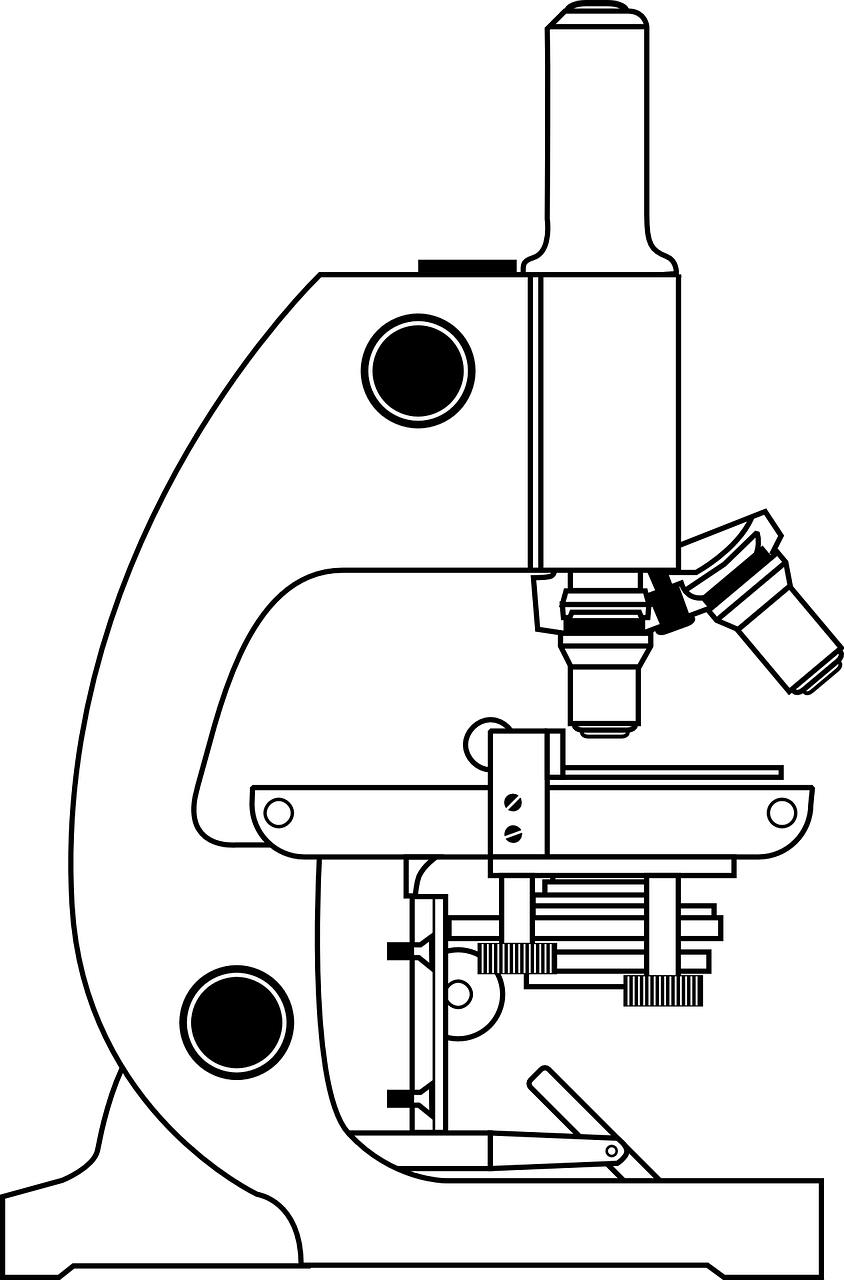
43 blank microscope diagram
The Parts of a Microscope (Blank) Printable - TeacherVision The Parts of a Microscope (Labeled) Printable. This diagram labels and explains the function of each part of a microscope. Project-Based Learning. No prep, ready-to-teach PBL for science, math, ELA, and social studies. Back to School 2022 Headquarters. Microscope Diagram Labeled, Unlabeled and Blank - Pinterest Sep 29, 2020 - Print a microscope diagram, microscope worksheet, or practice microscope quiz in order to learn all the parts of a microscope.
Microscope Diagram - Fill and Sign Printable Template Online | US Legal ... The tips below will allow you to complete Microscope Diagram quickly and easily: Open the template in the full-fledged online editor by hitting Get form. Fill out the necessary fields which are colored in yellow. Click the green arrow with the inscription Next to move from field to field. Go to the e-signature tool to add an electronic ...

Blank microscope diagram
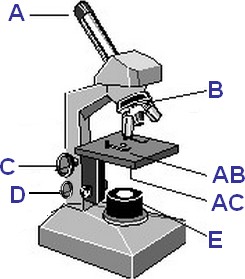
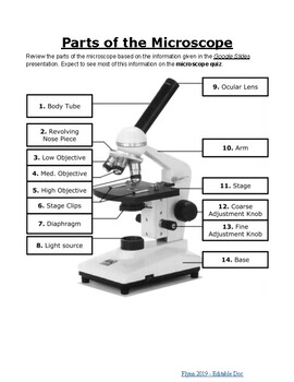
Microscope Labeling - The Biology Corner 1) Start with scanning (the shortest objective) and only use the COARSE knob . Once it is focused… 2) Switch to low power (medium) and only use the COARSE knob . You may need to recenter your slide. Once it is focused.. 3) Switch to high power (long objective). PDF Basic Microscopy Laboratory Exercises - Centers for Disease Control and ... 1. Correctly identify various parts of a brightfield microscope. Exercises: 1. Label the correct parts of a brightfield microscope on the graphic on the following page. 2. Identify the following parts of a brightfield microscope on the bench microscope you are using: A. Objectives B. Condenser (Iris) diaphragm C. Coarse adjustment Parts of the Microscope with Labeling (also Free Printouts) Microscopes are specially created to magnify the image of the subject being studied. This exercise is created to be used in homes and schools. the microscope layout, including the blank and answered versions are available as pdf downloads. Click to Download : Label the Parts of the Microscope (A4) PDF print version.
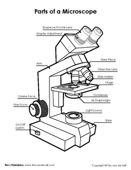
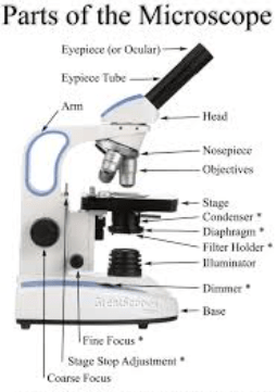
Blank microscope diagram. Microscope Labeling Game - Purpose Games This online quiz is called Microscope Labeling Game science, microsope. Microscope Diagram Labeled, Unlabeled and Blank | Parts of a ... Parts of a Microscope ... 1. Eyepiece/Ocular Lens – The lens into which the user looks to see the specimen. ... 3. Arm – A supporting piece of the optical ... Free Microscope Worksheets for Simple Science Fun for Your Students Parts of a Microscope The first worksheet labels the different parts of a microscope, including the base, slide holder, and condenser. I f you have a microscope, compare and contrast this worksheet to it. Also, your kids can color this microscope diagram in and read the words to each part of the microscope. Compound Microscope Parts, Functions, and Labeled Diagram Common compound microscope parts include: Eyepiece (ocular lens) with or without Pointer: The part that is looked through at the top of the compound microscope. Eyepieces typically have a magnification between 5x & 30x. Monocular or Binocular Head: Structural support that holds & connects the eyepieces to the objective lenses.
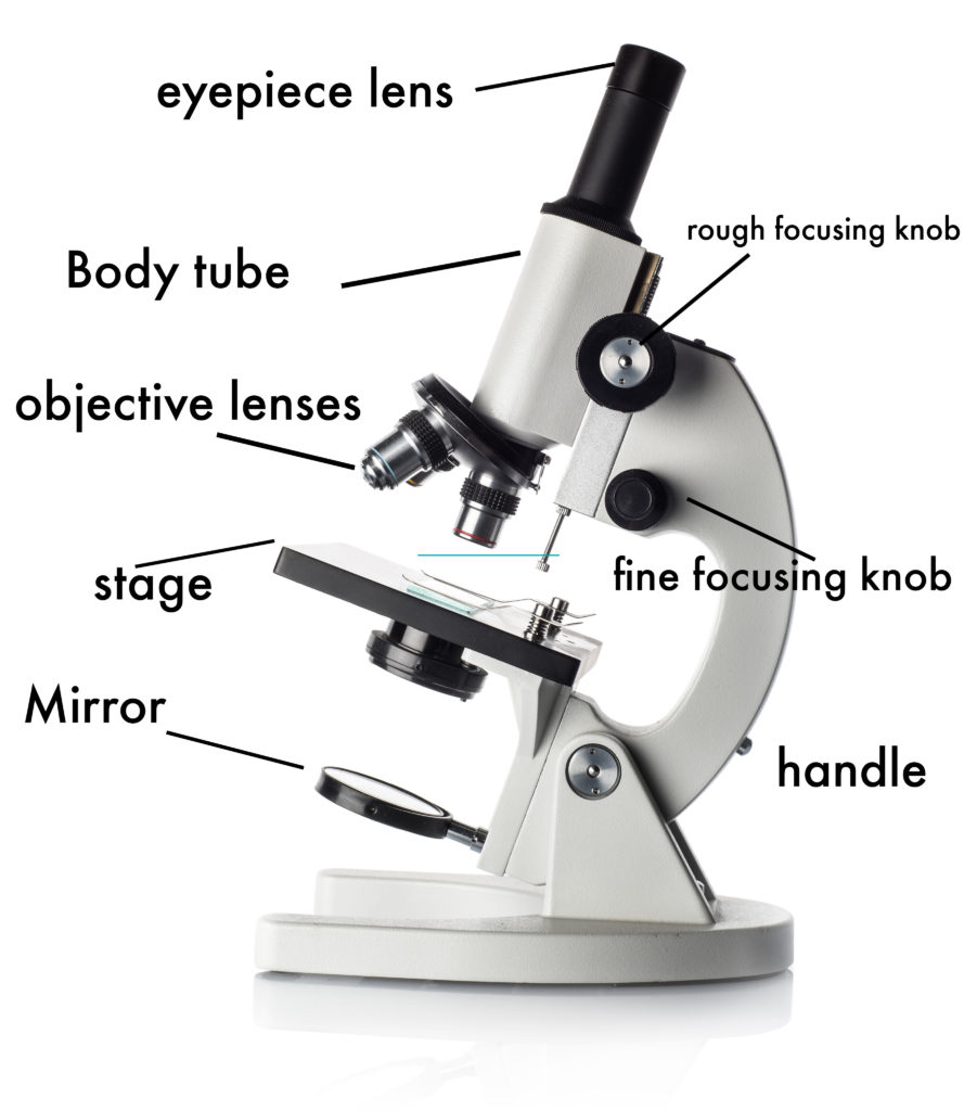
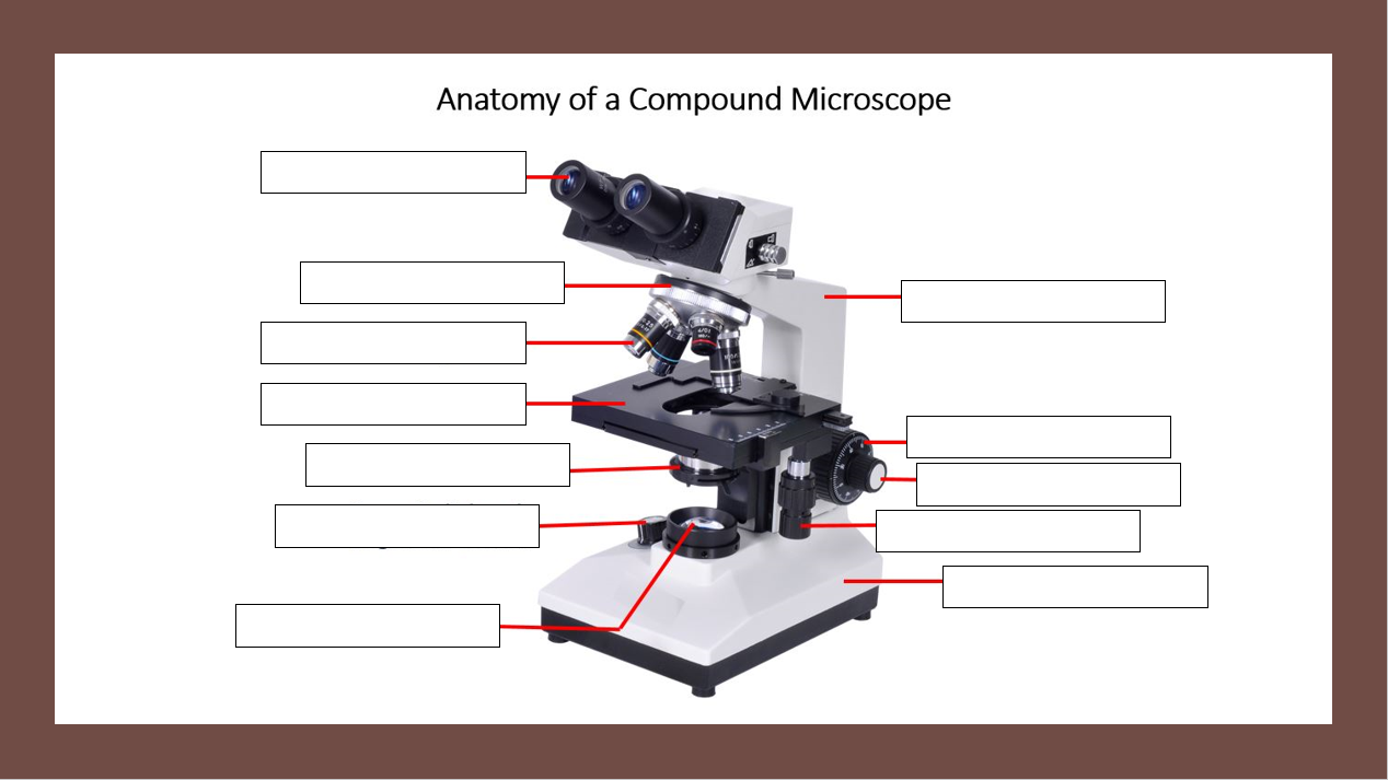
20 Color the Microscope Parts Worksheet | Worksheet From Home Microscope Parts Diagram PDF Science Printables color the microscope parts worksheet answers, color the microscope parts worksheet, via: timvandevall.com. Numbering Worksheets for Kids. Kids are usually introduced to this topic matter during their math education. The main reason behind this is that learning math can be done with the worksheets. Microscope Diagram - Tim's Printables Microscope Parts Diagram – Color, Labeled; Microscope Parts Diagram – Black and White, Labeled; Blank Microscope for Labeling Diagram – Color ... Clipart Panda - Free Clipart Images 50 images Blank Microscope Diagram Use these free images for your websites, art projects, reports, and Powerpoint presentations! Advertisement ©2020 ClipartPanda.com ... Microscope Parts & Functions - AmScope Head: The upper part of the microscope houses the eyepiece and objective lenses. Tube: Where the eyepieces are dropped in.Also, it connects the eyepieces to the objective lenses. Stage: The flat platform that supports the slides.Stage clips hold the slides in place. If your microscope has a mechanical stage, the slide is controlled by turning two knobs instead of having to move it manually.
Microscope Diagram Labeled, Unlabeled and Blank - Pinterest Dec 15, 2017 - Print a microscope diagram, microscope worksheet, or practice microscope quiz in order to learn all the parts of a microscope. Label the microscope — Science Learning Hub Interactive Label the microscope Interactive Add to collection Use this interactive to identify and label the main parts of a microscope. Drag and drop the text labels onto the microscope diagram. eye piece lens diaphragm or iris coarse focus adjustment stage base fine focus adjustment light source high-power objective Download Exercise Tweet 7th Microscope Diagram Worksheet-2 Microscope Worksheet. Use the words from this word list to identify the parts of the microscope. When you can identify a part of the microscope place the ... Microscope Parts and Functions First, the purpose of a microscope is to magnify a small object or to magnify the fine details of a larger object in order to examine minute specimens that cannot be seen by the naked eye. Here are the important compound microscope parts... Eyepiece: The lens the viewer looks through to see the specimen.
Parts of the Microscope with Labeling (also Free Printouts ...
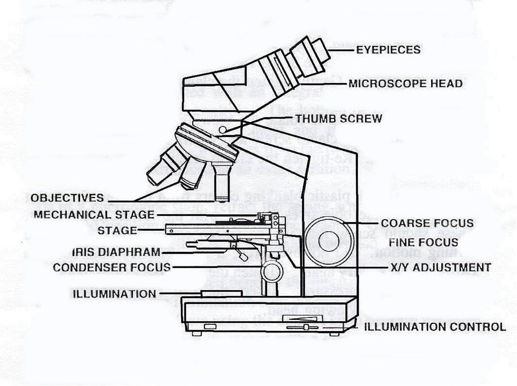
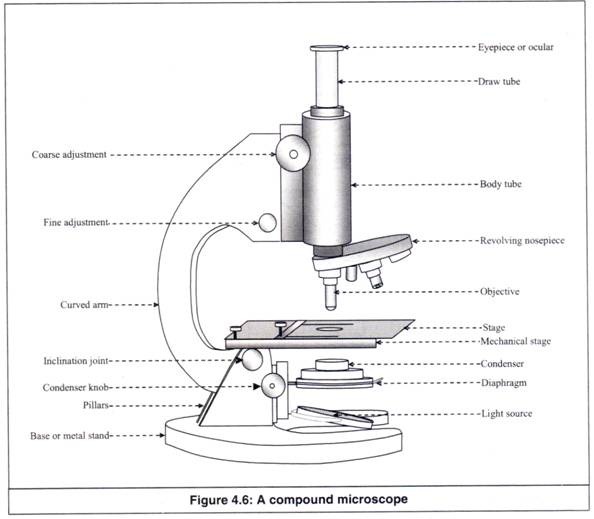
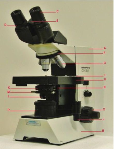
PDF The Microscope Parts and Use - Plainview the year 1590. The compound microscope uses lenses and light to enlarge the image and is also called an optical or light microscope (vs./ an electron microscope) . The simplest optical microscope is the magnifying glass and is good to about ten times (10X) magnification. The
Label a microscope - Teaching resources
16 Parts of a Compound Microscope: Diagrams and Video Once you have an understanding of the parts of the microscope it will be much easier to navigate around and begin observing your specimen, which is the fun part! The 16 core parts of a compound microscope are: Head (Body) Arm. Base. Eyepiece. Eyepiece tube.
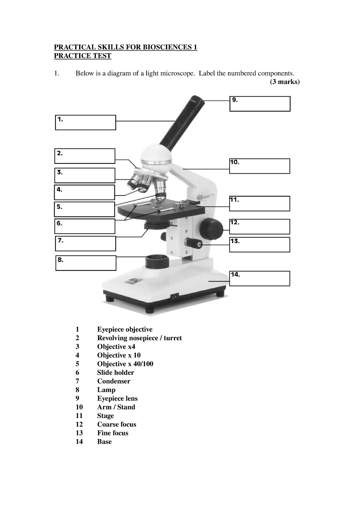
Practical Skills Spot Test Practice Answers - PRACTICAL ...
Microscope Labeling Diagram | Quizlet Focus and magnify light in differing amounts to view the specimen. Stage Clips. Hold the slide in place on the stage. Nosepiece. Holds the objective lenses and allows the lenses to rotate for viewing. Stage. Supports the slide where the specimen is being viewed. Lamp. Projects or reflects light upward through the diaphragm.
PRACTICAL BOOKLET - BIOLOGY4ISC
Microscope Diagram Labeled, Unlabeled and Blank | Parts of a Microscope ... timvandevall.com Microscope Diagram Labeled, Unlabeled and Blank | Parts of a Microscope Print a microscope diagram, microscope worksheet, or practice microscope quiz in order to learn all the parts of a microscope. Tim's Printables 39k followers More information Microscope Diagram Find this Pin and more on Science Printables by Tim's Printables.
Unit 1 How to Use Microscope
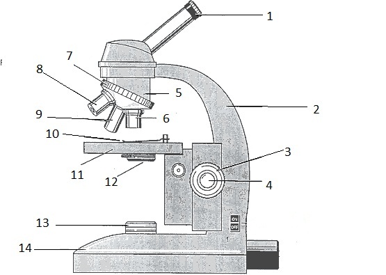
Microscope Labeling - The Biology Corner 18. You should carry the microscope by the _____ and the _____. 19. The objectives are attached to what part of the microscope (it can be rotated to click lenses into place?) _____ 20. A microscope has an ocular objective of 10x and a high power objective of 50x, what is the microscope's total magnification?
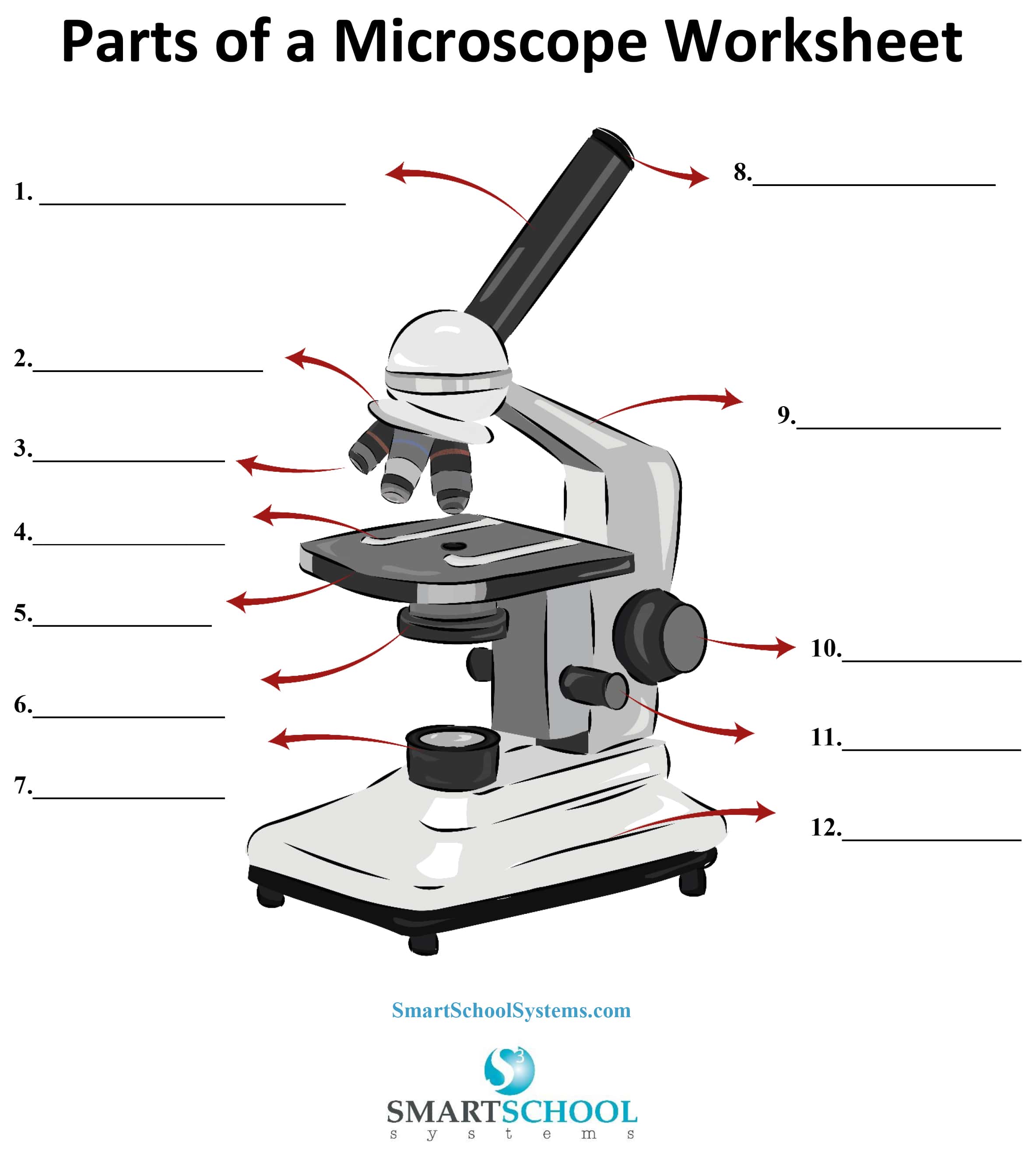
Parts of a Microscope - SmartSchool Systems
Simple Microscope - Parts, Functions, Diagram and Labelling Parts of the optical parts are as follows: Mirror - A simple microscope has a plano-convex mirror and its primary function is to focus the surrounding light on the object being examined. Lens - The biconvex lens is placed above the stage and its function is to magnify the size of the object being examined.
Parts of a Microscope - Free Printable
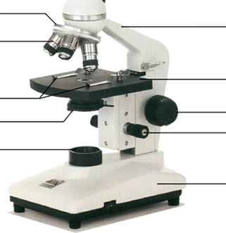
Microscope Diagram Teaching Resources | Teachers Pay Teachers Parts of a Microscope Overview Reading Comprehension and Diagram Worksheet by Teaching to the Middle 4.8 (24) $1.50 PDF This passage briefly describes microscopes and their parts (900-1000 Lexile). 14 questions (matching and multiple choice) assess students' understanding. Students label a diagram of 6 parts of a microscope.

Microscope for Kids, Dual Light Microscope Science Kit for Beginners Educational STEM Toy with 80X-1200X Magnification, Prepared and Blank Slides for ...
Learn About Microscopes With Fun, Free Printables - ThoughtCo Most microscopes used in a classroom setting are compound microscopes. These usually consist of a light source and three to five lenses with a total magnification of 40x to 1000x. The following free printables can help you teach your students the basic parts of a microscope so that they are ready to dive into a world previously unseen.
How to Use a Microscope
A Study of the Microscope and its Functions With a Labeled Diagram ... These labeled microscope diagrams and the functions of its various parts, attempt to simplify the microscope for you. However, as the saying goes, 'practice makes perfect', here is a blank compound microscope diagram and blank electron microscope diagram to label.

Compound and Stereo- microscopes - Microscopes 4 Schools
Microscope Diagram - Free Printable Tests and Worksheets | Biology ... When students map a microscope, they assign colors and/or patterns to each part they need to identify. They fill in the key and the corresponding part. This file includes two types of microscope maps: 1. a microscope with the twelve parts listed along the side 2. a microscope with arrows that allows the student to… J Jackie Schoen Teaching Things

Microscope Parts & Functions - AmScope
How to Sketch a Microscope Slide - Identifying and Sketching Cell ... Sketches come to life when you add highlights, shadows and color. For a pencil sketch, separate areas into white, light, medium and dark grey and black. To see the light/dark areas, squint so that the hard edges are blurred and your focus is on the shading. Start shading the light areas by following the shapes.

Label the microscope — Science Learning Hub
PDF Label parts of the Microscope Label parts of the Microscope: . Created Date: 20150715115425Z
blank diagram of a compound light microscope - Clip Art Library
Labeling the Parts of the Microscope | Microscope World Resources Labeling the Parts of the Microscope This activity has been designed for use in homes and schools. Each microscope layout (both blank and the version with answers) are available as PDF downloads. You can view a more in-depth review of each part of the microscope here. Download the Label the Parts of the Microscope PDF printable version here.

The Compound Microscope parts & how ... | Microscope parts ...
Diagram of a Compound Microscope - Biology Discussion 1. It is noted first that which objective lens is in use on the microscope. 2. Stage micrometer is positioned in such a way that it is in the field of view. 3. The eyepiece is rotated so that the two scales, the eyepiece or ocular scale and the stage micrometer scale, are parallel. 4.

Types of Microscopes: Definition, Working Principle, Diagram ...
Microscope: Types of Microscope, Parts, Uses, Diagram - Embibe There microscope anatomy includes three structural parts, i.e. head, base, and arm. Head - This is also known as the body; it carries the optical parts in the upper part of the microscope.. Base - It acts as microscopes support.It also carries microscopic illuminators. Arms - The microscope arm connects the base and the head and the eyepiece tube to the microscope base.
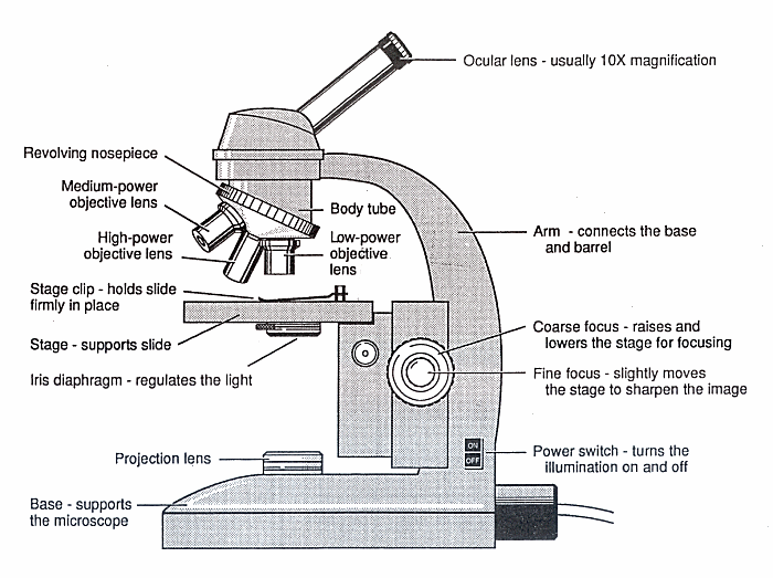
Diagram of a Microscope - Guide to using a microscope
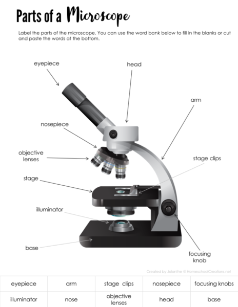
PDF Parts of a Microscope Printables - Homeschool Creations The lenses in a microscope make items appear smaller. How many parts of a microscope can you identify? Can you show the arm, stage, eyepiece, head, objective lens, illuminator, nosepiece, and stage clips? Where is the safest place to hold or carry a microscope? Which part of the microscope holds the specimen slide in place? Why do we use ...
(159).jpg)
Microscope Quiz: How Much You Know About Microscope Parts And ...
Parts of a microscope with functions and labeled diagram - Microbe Notes Figure: Diagram of parts of a microscope There are three structural parts of the microscope i.e. head, base, and arm. Head - This is also known as the body. It carries the optical parts in the upper part of the microscope. Base - It acts as microscopes support. It also carries microscopic illuminators.
Introduction to the Microscope Lab Activity
Parts of the Microscope with Labeling (also Free Printouts) Microscopes are specially created to magnify the image of the subject being studied. This exercise is created to be used in homes and schools. the microscope layout, including the blank and answered versions are available as pdf downloads. Click to Download : Label the Parts of the Microscope (A4) PDF print version.
Microscope Diagram and Quiz
PDF Basic Microscopy Laboratory Exercises - Centers for Disease Control and ... 1. Correctly identify various parts of a brightfield microscope. Exercises: 1. Label the correct parts of a brightfield microscope on the graphic on the following page. 2. Identify the following parts of a brightfield microscope on the bench microscope you are using: A. Objectives B. Condenser (Iris) diaphragm C. Coarse adjustment
Microscope Quiz
Microscope Labeling - The Biology Corner 1) Start with scanning (the shortest objective) and only use the COARSE knob . Once it is focused… 2) Switch to low power (medium) and only use the COARSE knob . You may need to recenter your slide. Once it is focused.. 3) Switch to high power (long objective).

Unlabeled microscope diagram | Clipart Panda - Free Clipart ...

Compound Microscope Parts – Labeled Diagram and their ...

Microscope Label Diagram | Quizlet
Human Anatomy
Microscope Parts Quiz
PARTS AND FUNCTIONS OF A COMPOUND MICROSCOPE

easy compound microscope diagram - Clip Art Library

Microscope Diagram - Free Printable Tests and Worksheets ...
Parts of the Microscope Labeling Activity!

parts of the microscope

AmScope B690B Siedentopf Binocular Compound Microscope, 40X-2000X Magnification, WH10x and WH20x Super-Widefield Eyepieces, Infinity Objectives, ...

The Compound Microscope Diagram | Quizlet

Living Environment Course
microscope vector sketch 7312859 Vector Art at Vecteezy
Microscope 40X / 100X / 400X / 1000X (Model 3000F-100-LED)

How to draw compound of Microscope easily - step by step
Different Types of Microscopes and their parts and function ...
Click and Learn - The Microscope - Parts
Microscopes - SCIENTIST CINDY

Microscope Components - Science Quiz

The Parts of a Microscope Quiz

Label Microscope Diagram - EnchantedLearning.com
Parts of a Microscope


Komentar
Posting Komentar