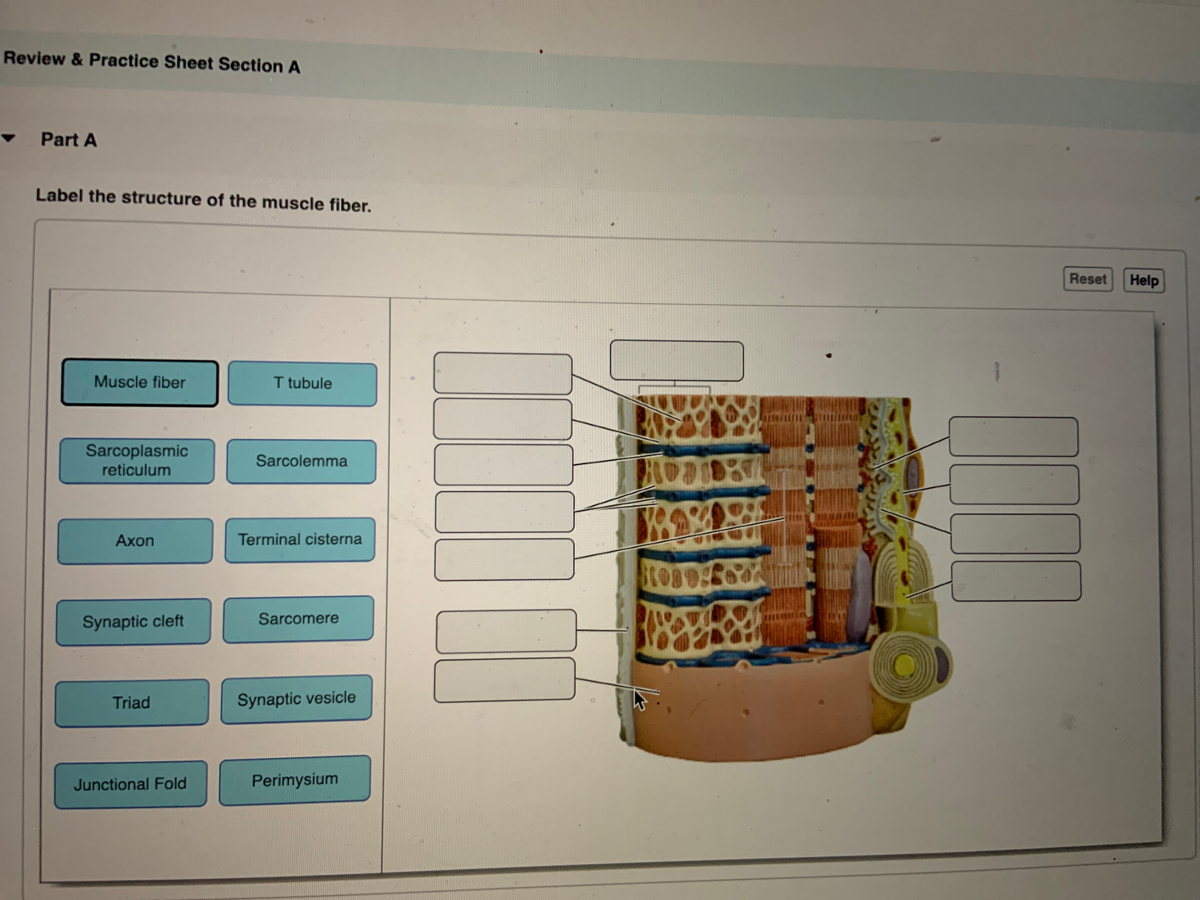
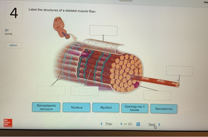
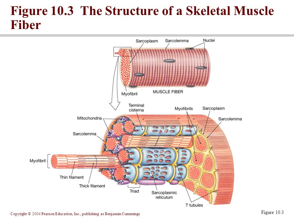
44 label the structures of a skeletal muscle fiber.
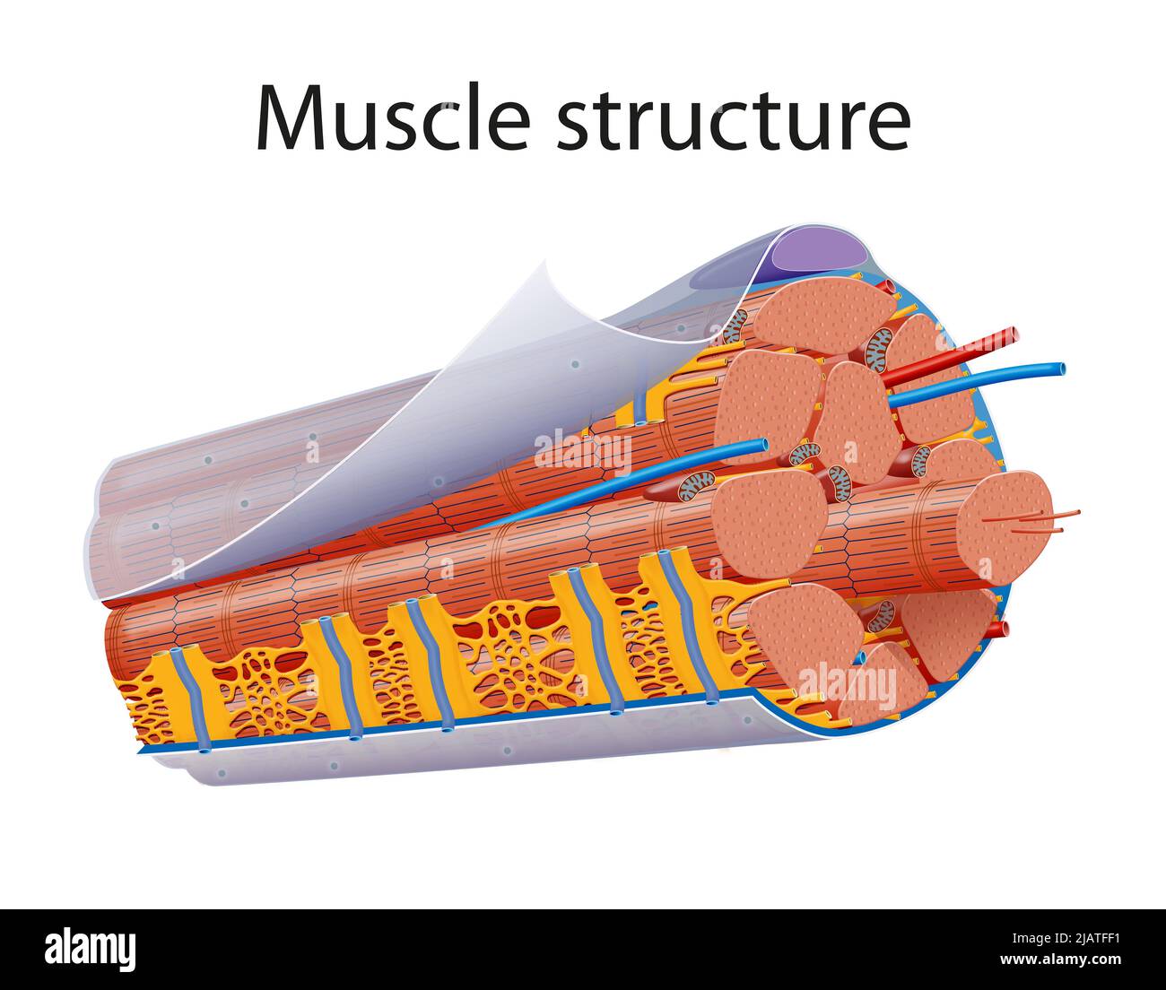
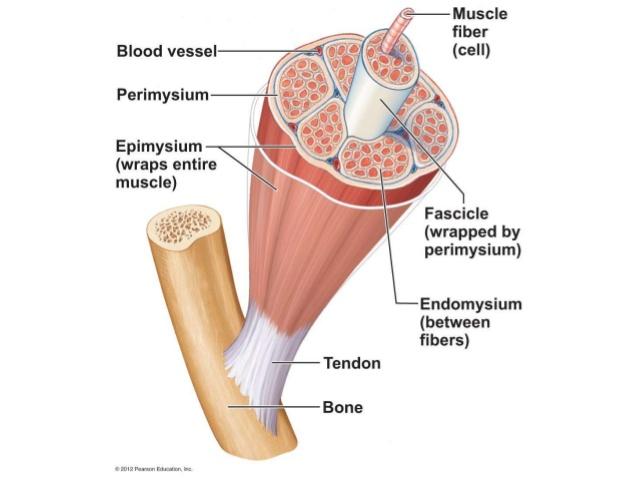
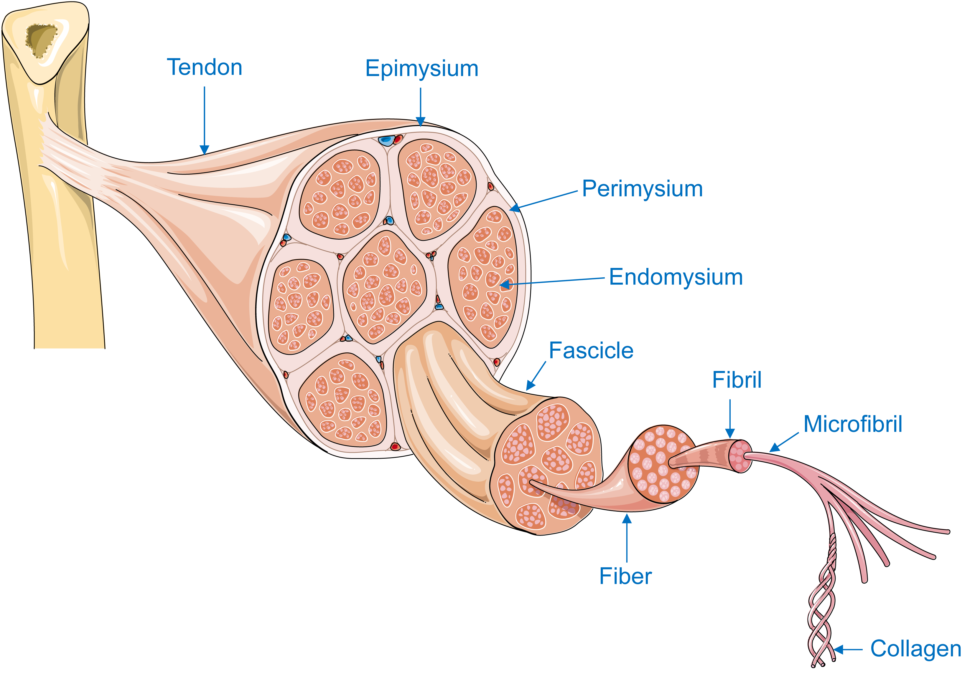
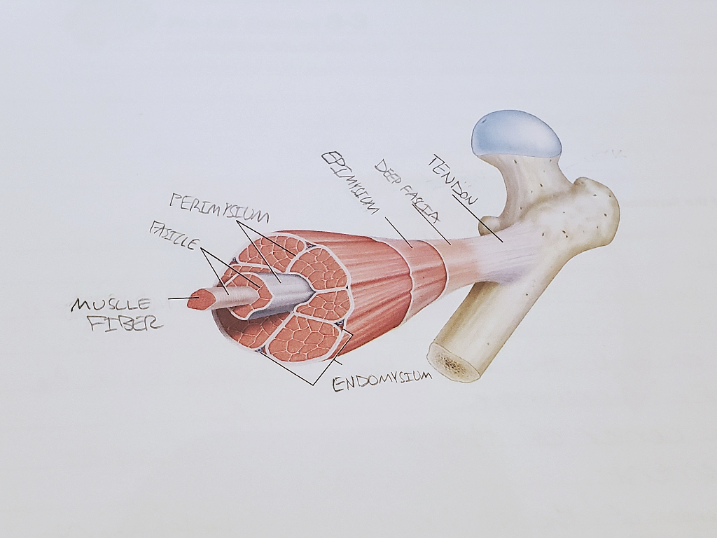
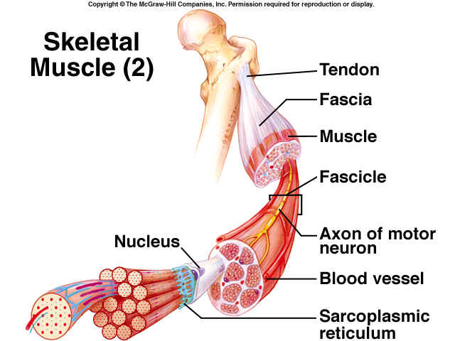
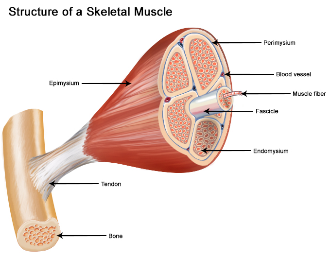
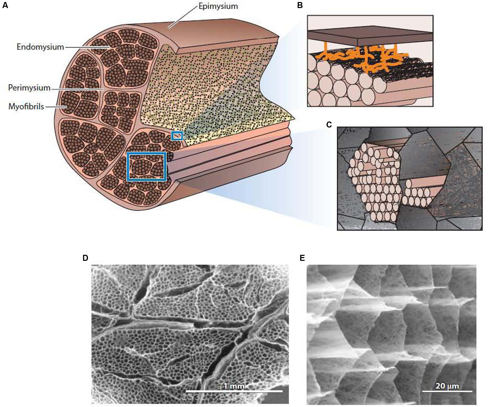
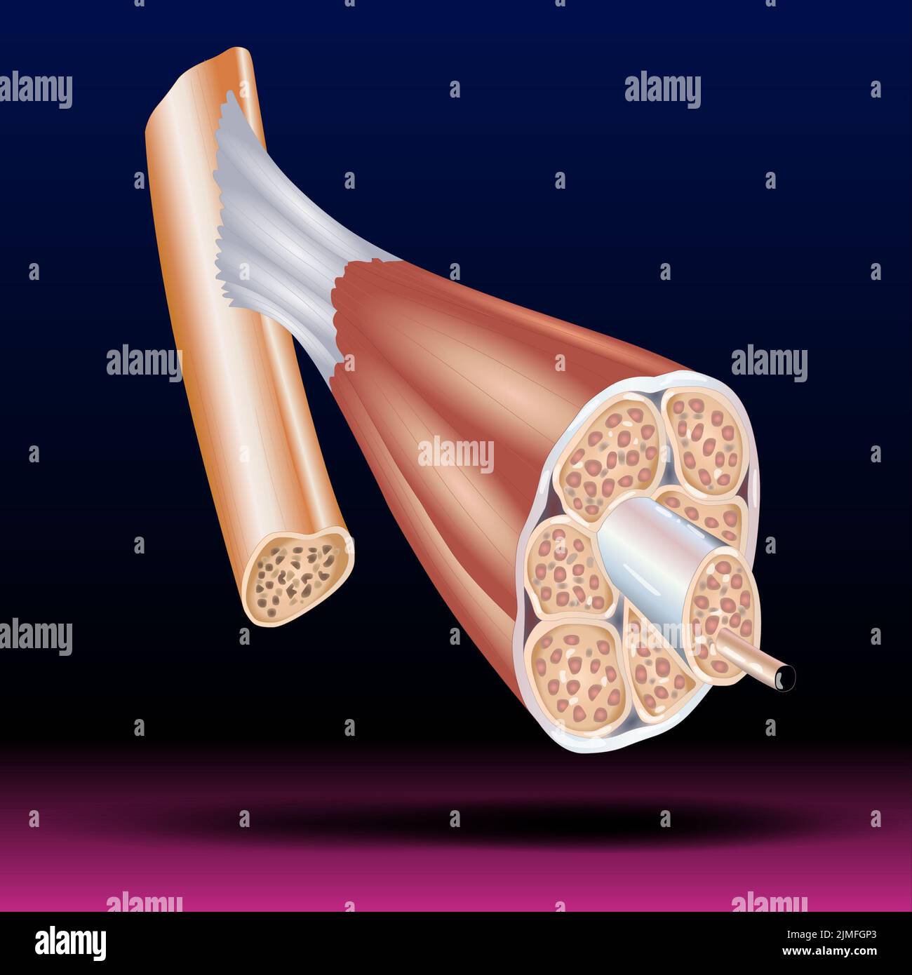
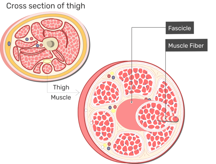
10.2 Skeletal Muscle – Anatomy & Physiology Figure 10.2.1 – The Three Connective Tissue Layers: Bundles of muscle fibers, called fascicles, are covered by the perimysium. Muscle fibers are covered by the endomysium. Inside each skeletal muscle, muscle fibers are organized into bundles, called fascicles, surrounded by a middle layer of connective tissue called the perimysium. 9.2A: Skeletal Muscle Fibers - Medicine LibreTexts Jan 17, 2023 · Skeletal Muscle Fiber Structure. Myocytes, sometimes called muscle fibers, form the bulk of muscle tissue. They are bound together by perimysium, a sheath of connective tissue, into bundles called fascicles, which are in turn bundled together to form muscle tissue. Myocytes contain numerous specialized cellular structures which facilitate their ...
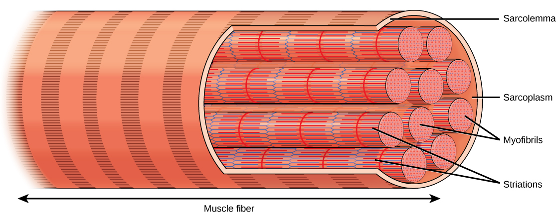
Skeletal Muscle | Anatomy and Physiology I - Lumen Learning Because skeletal muscle cells are long and cylindrical, they are commonly referred to as muscle fibers. Skeletal muscle fibers can be quite large for human cells, with diameters up to 100 μm and lengths up to 30 cm (11.8 in) in the Sartorius of the upper leg. During early development, embryonic myoblasts, each with its own nucleus, fuse with up to ...
Label the structures of a skeletal muscle fiber.
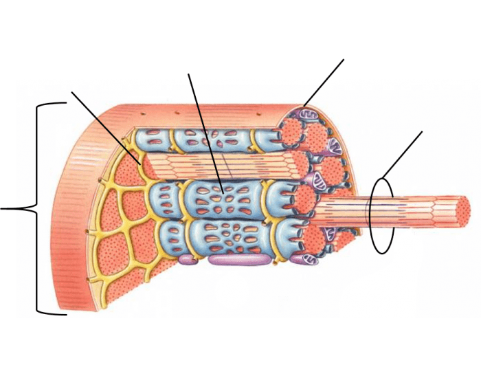
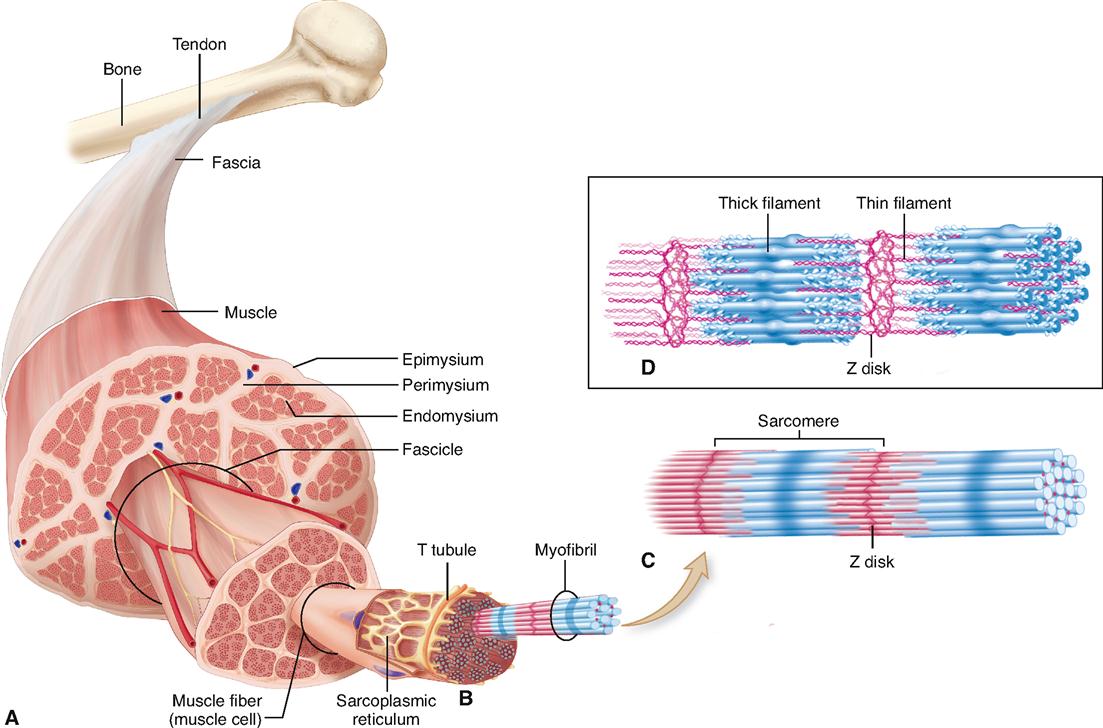
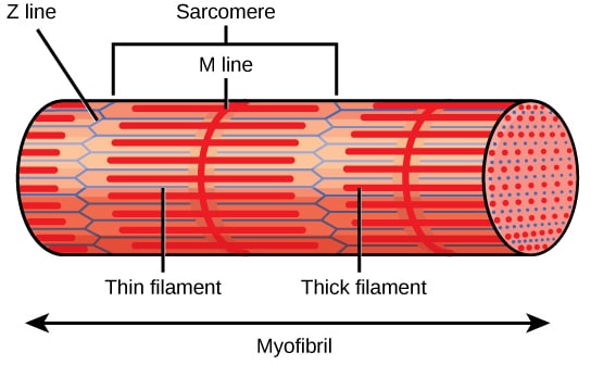
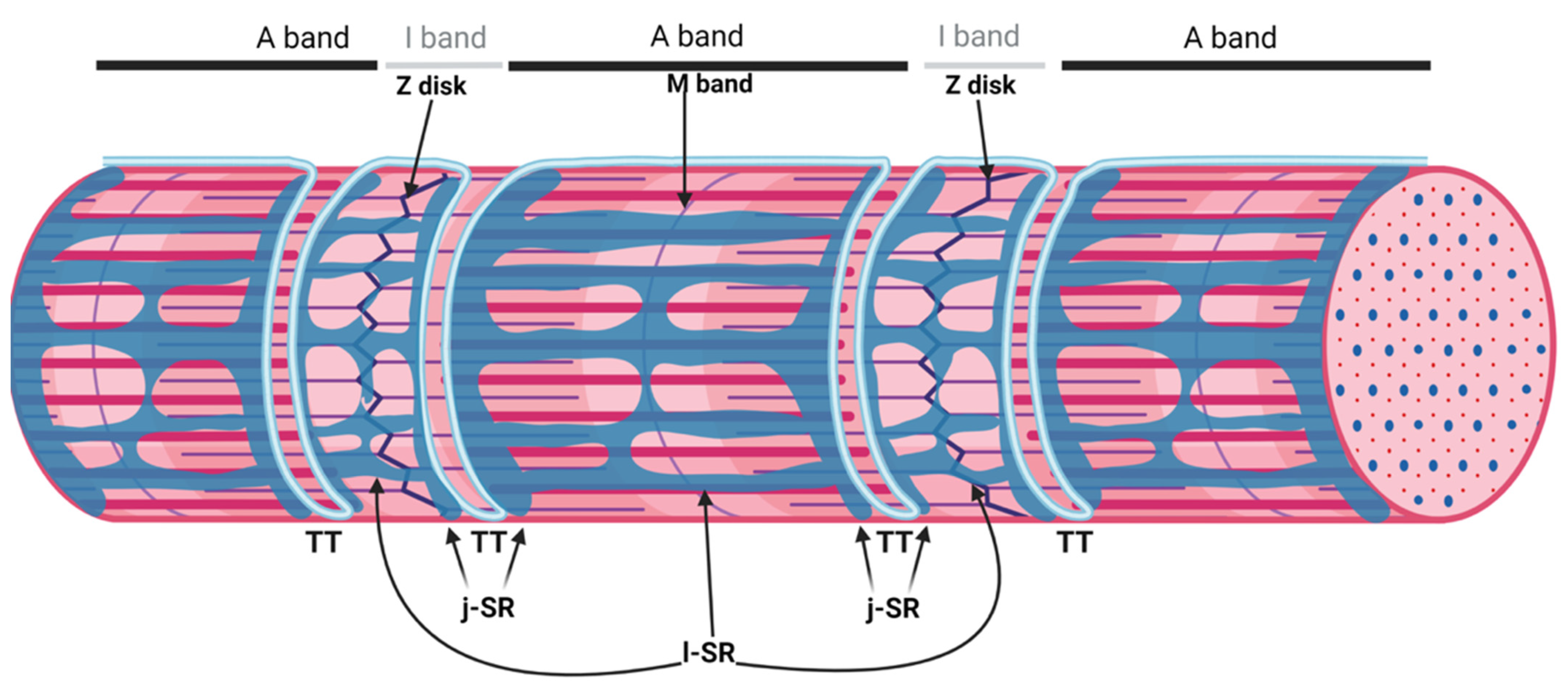
Muscle Fibers: Anatomy, Function, and More - Healthline May 12, 2020 · Skeletal muscle fibers are classified into two types: type 1 and type 2. Type 2 is further broken down into subtypes. Type 1. These fibers utilize oxygen to generate energy for movement. Type... Skeletal Muscle Fiber Structure and Function | Open Textbooks ... Apr 6, 2016 · The striated appearance of skeletal muscle tissue is a result of repeating bands of the proteins actin and myosin that occur along the length of myofibrils. Myofibrils are composed of smaller structures called myofilaments. There are two main types of myofilaments: thick filaments and thin filaments. Label structure of skeletal muscle Diagram | Quizlet Label structure of skeletal muscle 4.0 (5 reviews) + − Learn Test Match Created by danielaaaa04 Terms in this set (8) myofibrils ... sarcoplasmis reticulum ... sarcolemma ... epimysium ... perimysium ... endomysium ... fascicle ... muscle fiber ...
Label the structures of a skeletal muscle fiber.. Label structure of skeletal muscle Diagram | Quizlet Label structure of skeletal muscle 4.0 (5 reviews) + − Learn Test Match Created by danielaaaa04 Terms in this set (8) myofibrils ... sarcoplasmis reticulum ... sarcolemma ... epimysium ... perimysium ... endomysium ... fascicle ... muscle fiber ... Skeletal Muscle Fiber Structure and Function | Open Textbooks ... Apr 6, 2016 · The striated appearance of skeletal muscle tissue is a result of repeating bands of the proteins actin and myosin that occur along the length of myofibrils. Myofibrils are composed of smaller structures called myofilaments. There are two main types of myofilaments: thick filaments and thin filaments. Muscle Fibers: Anatomy, Function, and More - Healthline May 12, 2020 · Skeletal muscle fibers are classified into two types: type 1 and type 2. Type 2 is further broken down into subtypes. Type 1. These fibers utilize oxygen to generate energy for movement. Type...
Skeletal muscle hi-res stock photography and images - Alamy

Muscle Fiber (3B) - Pierce College - Anatomy

Solved ge Home Topic Application Technolog.. Seved 19 Label ...

Ch 10 lab map Flashcards | Quizlet
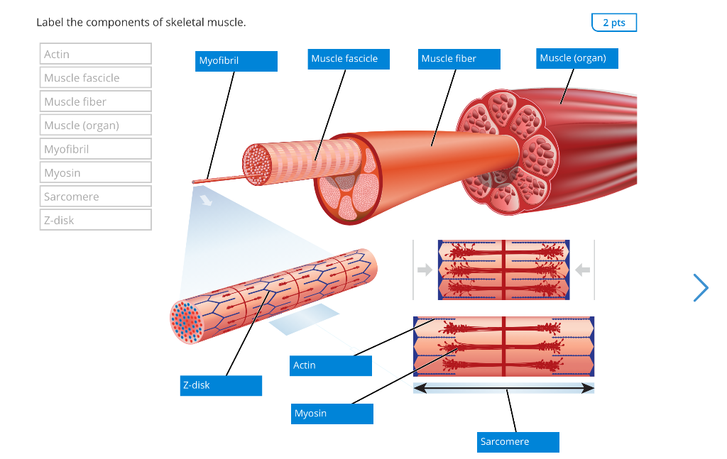
Solved Label the components of skeletal muscle. 2 pts Actin ...
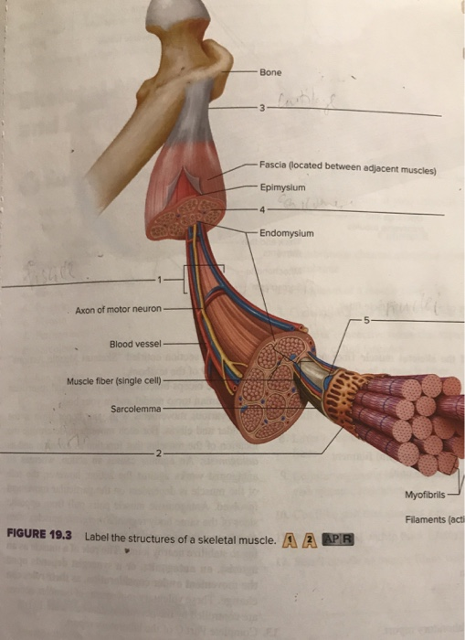
Solved Bone Fascia (located between adjacent muscles) | Chegg.com

Connect Exam 2 Flashcards | Quizlet

Bellringer 12/9 Label #4 and #5. Building Skeletal Muscle… We ...
Bio 201 Muscular System Flashcards - Easy Notecards
Servier - Drawing Structure of the skeletal muscle - English ...
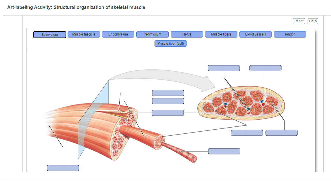
Answered: Art-labeling Activity: Structural… | bartleby
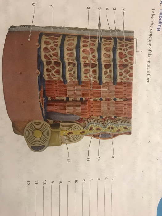
Solved A. Labeling Label the structure of the muscle fiber ...
Structure of a Skeletal Muscle Fiber Quiz
Solved Labeling the basic structures of skeletal muscles ...
Biology, Animal Structure and Function, The Musculoskeletal ...
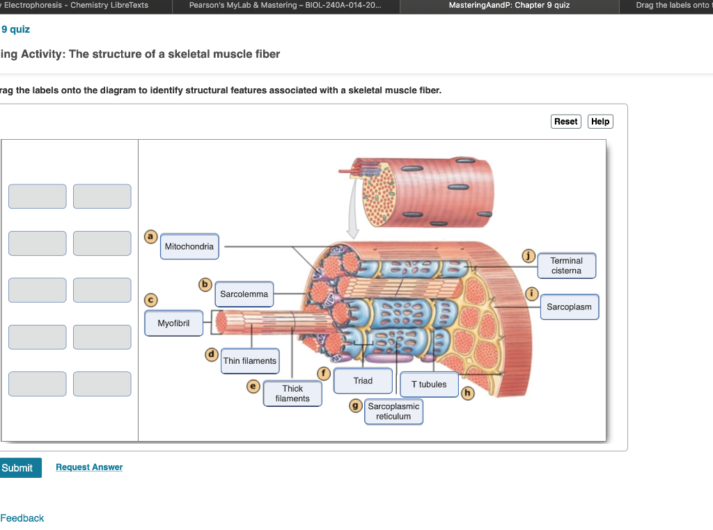
Solved Electrophoresis - Chemistry Libre Texts Pearson's ...
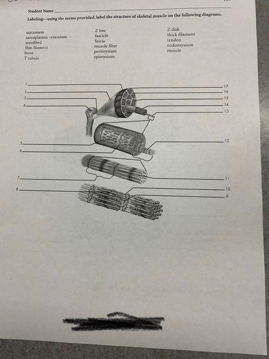
Solved Student Name Labeling-using the terms provided, label ...
Answered: Label the structure of the muscle fiber | bartleby

Solved Muscle Cell Label the structures of a skeletal muscle ...

ANSWER THIS NOW!!!! 40 points!!! Drag each label to the ...

Label the following in a diagram of a skeletal muscle fiber ...
The Muscular System

Draw a neuromuscular junction and label every structure ...
Physiology of the Muscular System | Basicmedical Key
Structure of Skeletal Muscle. | Download Scientific Diagram
SEER Training: Structure of Skeletal Muscle

ch 10 connect AP 150 Flashcards | Quizlet
Solved Hel Label the structures of a skeletal muscle fiber ...
Frontiers | The Structure and Role of Intramuscular ...
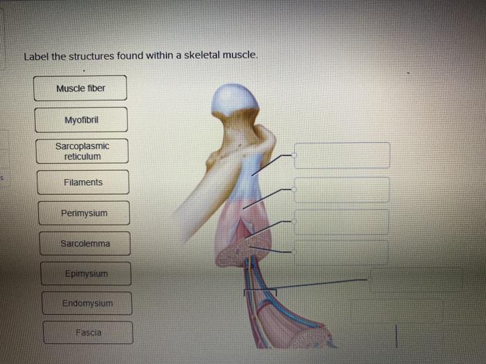
Solved Label the structures found within a skeletal muscle ...

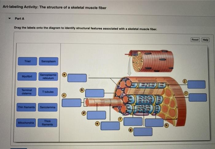
Art-labeling Activity: The Structure of a Skeletal Muscle ...

A) Illustration of skeletal muscle structure copied with ...

Triad (anatomy) - Wikipedia
Human Biology fig. 1.20 - Structure of a skeletal muscle ...
Solved Art-labeling Activity: The Structure of a Sarcomere ...
Endomysium hi-res stock photography and images - Alamy
Skeletal muscle - Structure - Contraction - TeachMePhysiology
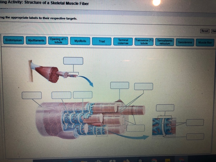
Solved -ling Activity: Structure of a Skeletal Muscle Fiber ...

skeletal muscle fiber labeled Diagram | Quizlet
Muscle Tissue. - ppt download

Label structure of skeletal muscle Diagram | Quizlet

Lab Practical #2 Flashcards | Quizlet
Skeletal muscle fibers: arrangement and diagram | GetBodySmart
Biomolecules | Free Full-Text | The Sarcoplasmic Reticulum of ...


Komentar
Posting Komentar