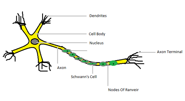
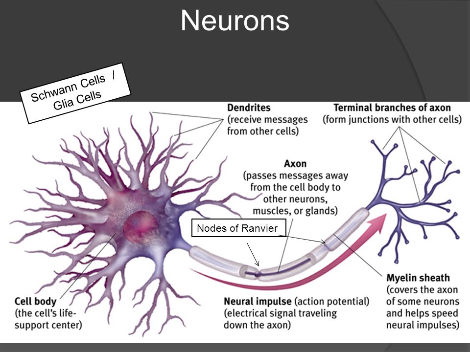
43 draw and label a neuron
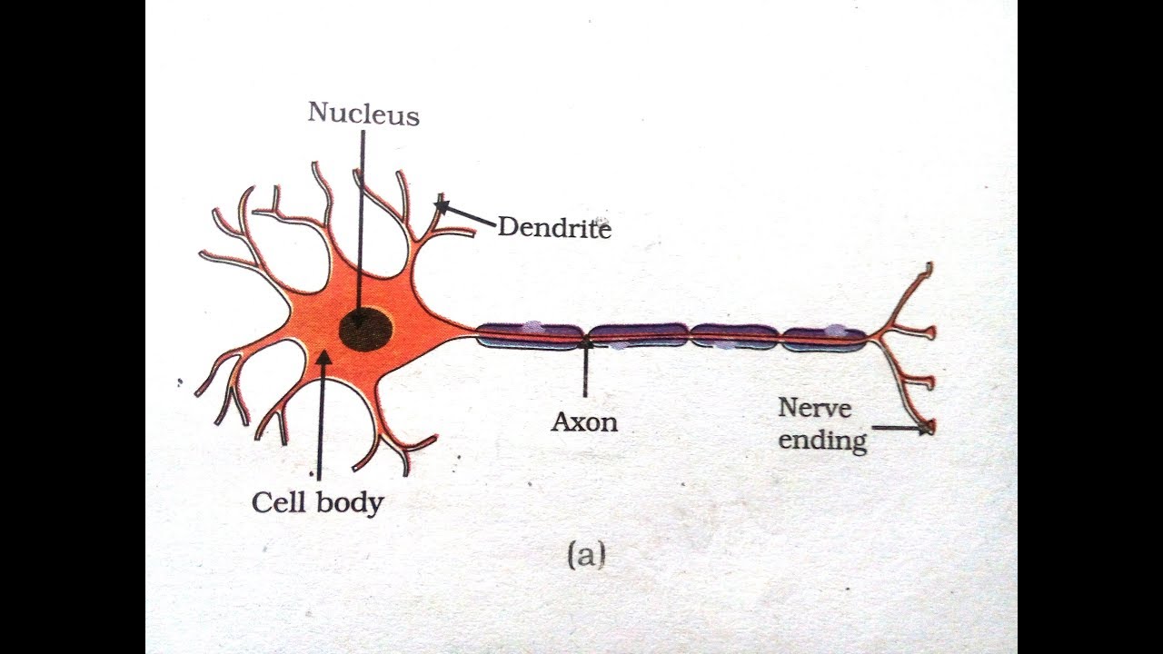
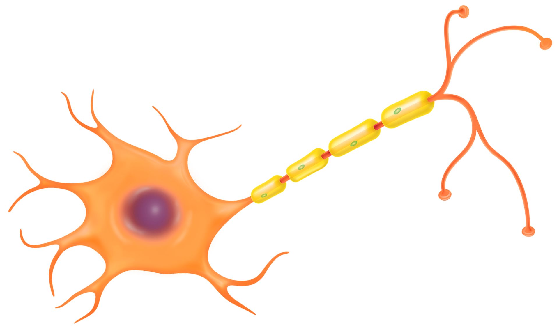
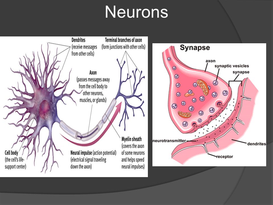
how to draw structure of neuron/neuron diagram labelled/diagram of ... Neuron Diagram Draw Diagram Body Diagram Neuron Structure And Function Cell Membrane Structure Plasma Membrane Biology Drawing Brain Drawing More information ... More information how to draw structure of neuron/neuron diagram labelled/diagram of neuron/neuron cell - YouTube Comments ermiradinho are these all the parts A anaroleon mui vien divujado Draw and label a neuron. Explain how it carries messages Answer: Neurons are the fundamental unit of the nervous system. Neurons are also called as nerve cells. The main function of the neurons is to pass the receiving information and send appropriate signals to the rest parts of the body. The signals received are in the form of electrical signals. Neuron comprises of dendrite, axon and cell body.
What Is a Neuron? - Definition, Structure, Parts and Function - BYJUS Neurons are the structural and functional unit of the nervous system. All neurons have three different parts - dendrites, cell body and axon. The neuron structure is specially adapted to carry messages over large distances in the body quickly in the form of electrical signals. What are sensory neurons and motor neurons?

Draw and label a neuron
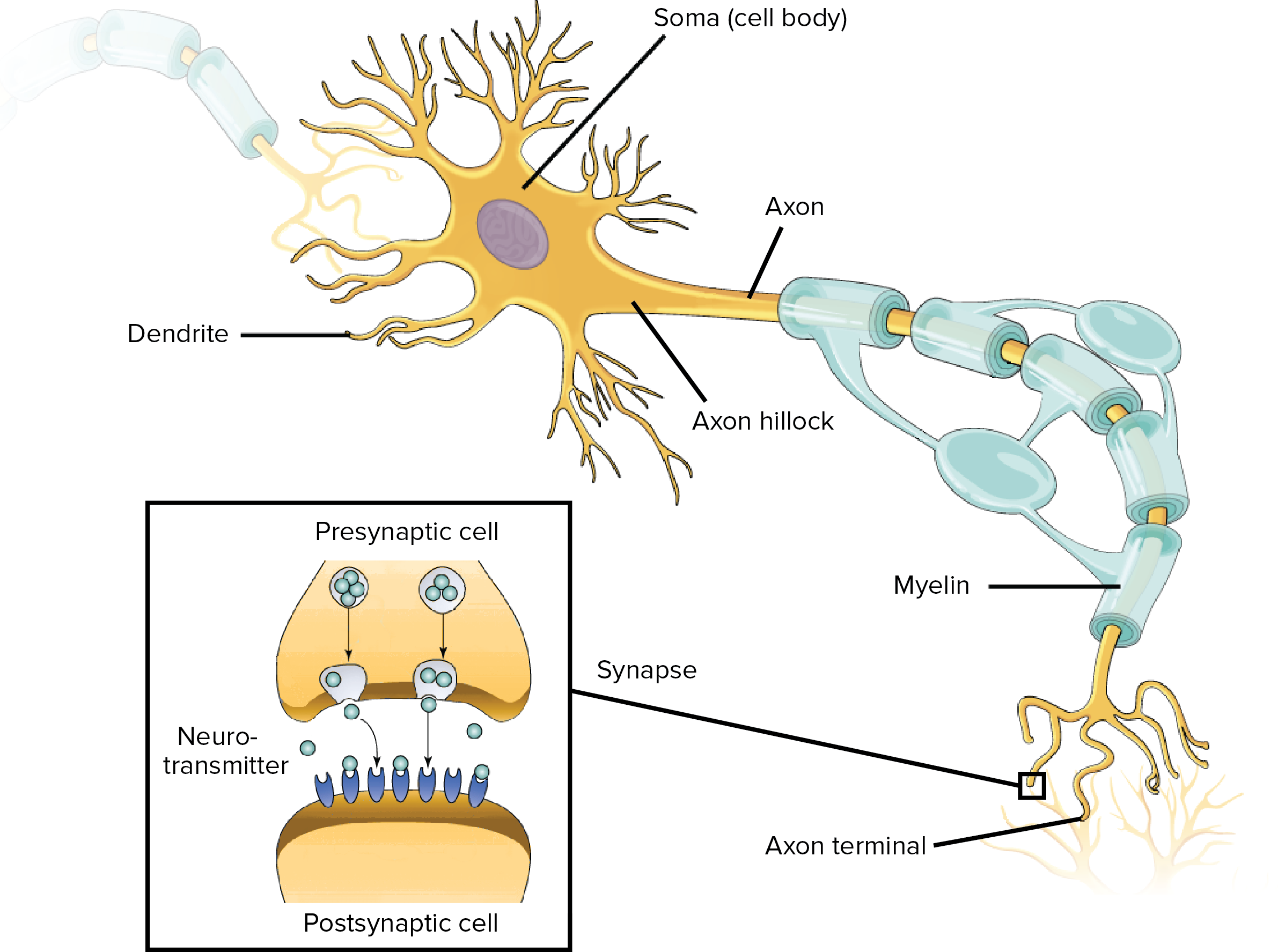
Diagram Quiz on Neuron Structure and Function (Labeling Quiz) 1. Identify the cell type in the above figure Liver Cell Cardiac Cell Nerve cell Skin cell. 2. In the figure, labeled '1' receives impulses from adjacent neuron. It is called the Dendron Dendrite Axon Axonite. 3. In the figure, labeled '2' is the short filaments from the cell body that carries impulses from dendrites to the cell body which is the Schwann cell Axonite Axon Dendron › science › biologyThe synapse (article) | Human biology | Khan Academy At a synapse, one neuron sends a message to a target neuron—another cell. Most synapses are chemical; these synapses communicate using chemical messengers. Other synapses are electrical; in these synapses, ions flow directly between cells. At a chemical synapse, an action potential triggers the presynaptic neuron to release neurotransmitters. Overview of neuron structure and function - Khan Academy Neurons are the basic functional units of the nervous system, and they generate electrical signals called action potentials, which allow them to quickly transmit information over long distances. Glia are also essential to nervous system function, but they work mostly by supporting the neurons.
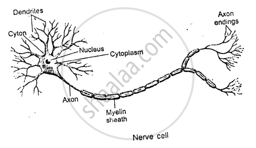
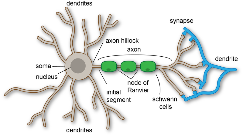
Draw and label a neuron. Nervous System - Label the Neuron - TheInspiredInstructor.com Nervous System - Neuron: Nerve Cell Name: Choose the correct names for the parts of the neuron. (1) (2) (3) (4) (5) (6) This neuron part receives messages from other neurons. (7) This neuron part sends on messages to other neurons. (8) This neuron part gives messages to muscle tissue. (9) This neuron part processes incoming messages. quizlet.com › 241571764 › label-parts-of-a-neuron-diagramLabel Parts of a Neuron Diagram | Quizlet Label Parts of a Neuron 4.2 (13 reviews) + − Flashcards Learn Test Match Created by cottonje Terms in this set (14) Dendrites receives impulses from other nerve cells axon hillock The cell body...the part of the cell that houses the nucleus and keeps the entire cell alive and functioning Myelin Sheath Draw a labelled diagram of the neuron and describe the structure of ... Draw a labelled diagram of the neuron and describe the structure of neuron in detail. Medium Solution Verified by Toppr The structure of neuron: Nerve cells or neurons are the structural and functional units of the nervous system. It consists of three major parts namely, Cell body, dendrites, Axon. Cell Body: It is irregular in shape or polyhedral. How to draw and label neuron | step by step tutorial - YouTube ... Jun 3, 2019 - A beautiful drawing of a Neuron. And it will teach you to draw the Neuron very easily. Watch the video and please be kind enough to thumbs up my videos. And ... Pinterest. Today. Watch. Explore. When autocomplete results are available use up and down arrows to review and enter to select. Touch device users, explore by touch or ...
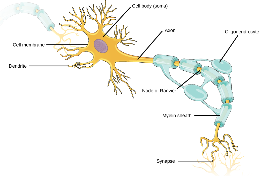
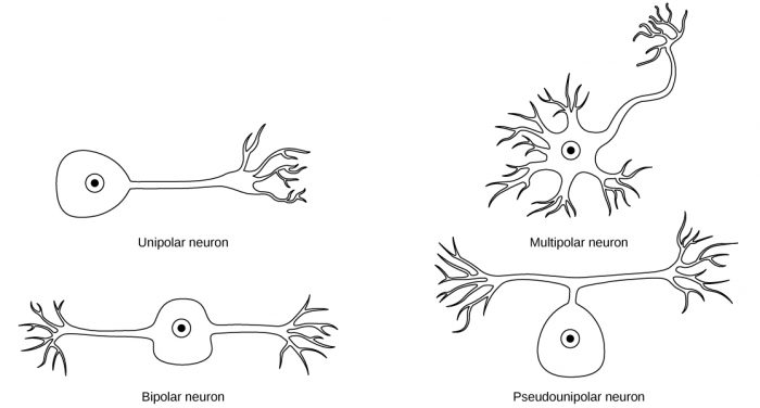
A Labelled Diagram Of Neuron with Detailed Explanations - BYJUS A neuron is also known as the nerve cell. The structure of a neuron varies with their shape and size and it mainly depends upon their functions and their location. Neurons are the structural and functional units of the nervous system. A group of neurons forms a nerve. Neurons are the structural and functional units of the nervous system. An Easy Guide to Neuron Anatomy with Diagrams - SimplyPsychology.org Neurons are the information processing units of the brain which have a responsibility for sending, receiving, and transmitting electrochemical signals throughout the body. Neurons, also known as nerve cells, are essentially the cells that make up the brain and the nervous system. Neurons do not touch each other, but where one neuron comes close ... NEURON STRUCTURE AND CLASSIFICATION - Brigham Young University-Idaho Structural classification of neurons. 1) Bipolar; 2) Multipolar and 3) Unipolar. Bipolar neurons have only two processes that extend in opposite directions from the cell body. One process is called a dendrite, and another process is called the axon. Although rare, these are found in the retina of the eye and the olfactory system. sciencetrends.com › labeled-neuron-diagramLabeled Neuron Diagram - Science Trends May 29, 2019 · Neurons are a type of cell and are the fundamental constituents of the nervous system and brain. Neurons take in stimuli and convert them to electrical and chemical signals that are sent to our brain. There are 3 major kinds of neurons in the spinal cord: sensory, motor, and interneurons.
Label Neuron Anatomy Printout - EnchantedLearning.com Label Neuron Anatomy Printout - EnchantedLearning.com Advertisement. EnchantedLearning.com is a user-supported site. As a bonus, site members have access to a banner-ad-free version of the site, with print-friendly pages. Click here to learn more. (Already a member? Click here.) You might also like: The Spinal Cord Brain Cells Brain Glossary Neuron Diagram || Diagram Of A Neuron || How To Draw A Neuron Step By ... Neuron Diagram || Diagram Of A Neuron || How To Draw A Neuron Step By Step For Beginners - YouTube 0:00 / 6:08 Neuron Diagram || Diagram Of A Neuron || How To Draw A Neuron Step By... Parts of a Neuron and How Signals are Transmitted - Verywell Mind Neurons are the basic building blocks of the nervous system. These specialized cells are the information-processing units of the brain responsible for receiving and transmitting information. Each part of the neuron plays a role in communicating information throughout the body. › watchHow To Draw A Neuron Step By Step For Beginners - YouTube How To Draw A Neuron Step By Step For Beginners Adimu Show 31.8K subscribers Subscribe 32K views 3 years ago A beautiful drawing of a Neuron. And it will teach you to draw the Neuron...
Neuron Label Diagram | Quizlet
Overview of neuron structure and function - Khan Academy Neurons are the basic functional units of the nervous system, and they generate electrical signals called action potentials, which allow them to quickly transmit information over long distances. Glia are also essential to nervous system function, but they work mostly by supporting the neurons.

Draw a neat and clean diagram of a neuron and label the ...
› science › biologyThe synapse (article) | Human biology | Khan Academy At a synapse, one neuron sends a message to a target neuron—another cell. Most synapses are chemical; these synapses communicate using chemical messengers. Other synapses are electrical; in these synapses, ions flow directly between cells. At a chemical synapse, an action potential triggers the presynaptic neuron to release neurotransmitters.

6.5.2 Draw and label a diagram of the structure of a motor neuron
Diagram Quiz on Neuron Structure and Function (Labeling Quiz) 1. Identify the cell type in the above figure Liver Cell Cardiac Cell Nerve cell Skin cell. 2. In the figure, labeled '1' receives impulses from adjacent neuron. It is called the Dendron Dendrite Axon Axonite. 3. In the figure, labeled '2' is the short filaments from the cell body that carries impulses from dendrites to the cell body which is the Schwann cell Axonite Axon Dendron

File:Complete neuron cell diagram en.svg - Wikipedia

6.5 Nerves, Hormones and Homeostasis | BioNinja

Label Parts of a Neuron Diagram | Quizlet
Draw a neat labelled diagram of multipolar myelinated neuron ...

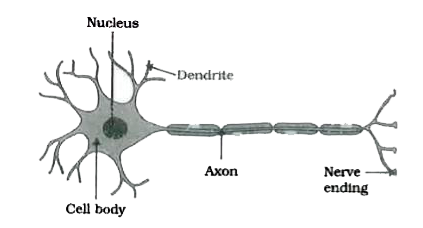
Draw nerve cell and label its parts - Brainly.in

Label Neuron Diagram | Quizlet
Draw and label the structure of a neuron. - Sarthaks eConnect ...

Draw the structure of a neuron and label its nucleus dendrite ...
How to draw a Motor Neuron

Draw and Label a Neuron. - Science | Shaalaa.com

Label the Parts of a Neuron | Neurons, Teaching biology ...

Neuron Diagram || Diagram Of A Neuron || How To Draw A Neuron Step By Step For Beginners

Draw a labelled diagram of a neuron.
Overview of neuron structure and function (article) | Khan ...

Draw and Label the Diagram. Nerve Cell - Science and ...
Draw a Labelled Diagram Showing the Structure of a Neuron ...
16.1 Neurons and Glial Cells – Concepts of Biology – 1st ...
Nerve Cell Function | Nerve Cell Diagram | DK Find Out

Draw the diagram of neuron and label any two parts.

Long answer question Draw the neat labelled diagram of ...
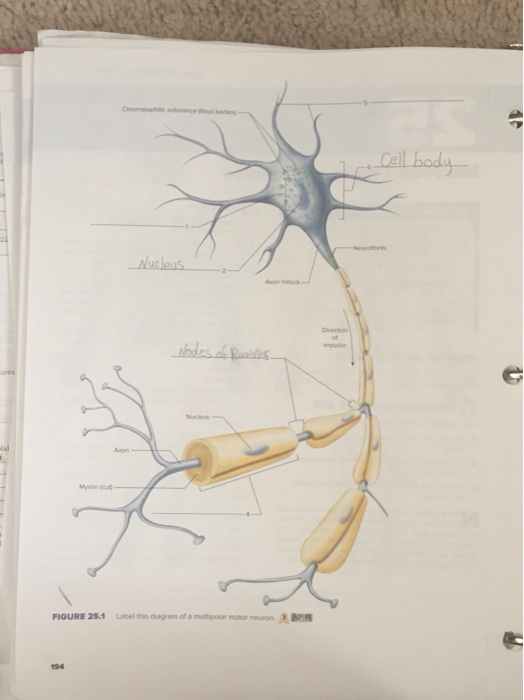
Solved ures Nucieus Axon FIQURE 25.1 Label this diagram of a ...

Draw and label the diagram of the nerve cell
Draw the structure of neuron and label cell body and axon.

What Is a Neuron? Diagrams, Types, Function, and More
Dendrite - Wikipedia

Draw neat labelled diagram of neuron.

Draw the structure of a neuron and label nucleus dendrite ...
Draw a neuron and a synapse and label as much of it as you ...

Draw a labelled diagram of a neuron
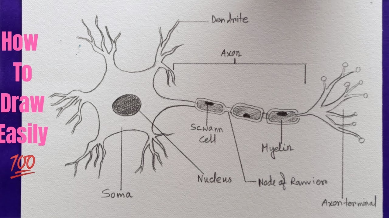
How to draw neuron diagram easily - step by step
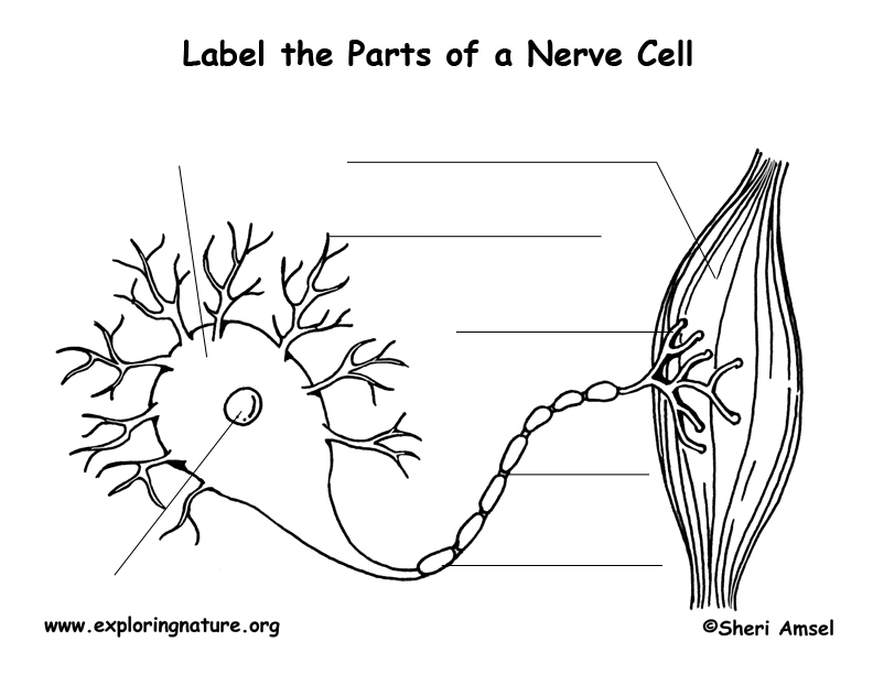
Nerve Cell (Neuron) Labeling Page

Q10. (a) Draw the structure of neuron and label cell body and ...

How to draw Neuron unit of nervous tissue | Structure of a ...
Labeled Neuron Diagram - Science Trends

Labeling the Neuron Diagram | Quizlet
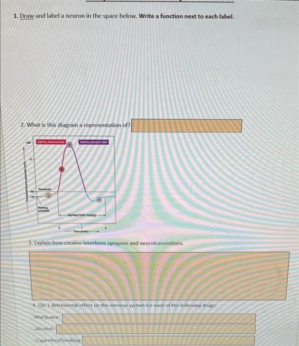
Solved 1. Draw and label a neuron in the space below. Write ...

Draw a neatly labelled diagram of a neuron. - Brainly.in

Draw the diagram of a neuron and label the following parts ...
Draw a neat labelled diagram of neurons.
Draw a neuron and a synapse and label as much of it as you ...


Komentar
Posting Komentar