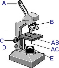
40 label the indicated parts of the microscope
Labeling the Parts of the Microscope | Microscope World Resources Labeling the Parts of the Microscope This activity has been designed for use in homes and schools. Each microscope layout (both blank and the version with answers) are available as PDF downloads. You can view a more in-depth review of each part of the microscope here. Download the Label the Parts of the Microscope PDF printable version here. Parts of a microscope with functions and labeled diagram - Microbe Notes Figure: Diagram of parts of a microscope There are three structural parts of the microscope i.e. head, base, and arm. Head - This is also known as the body. It carries the optical parts in the upper part of the microscope. Base - It acts as microscopes support. It also carries microscopic illuminators.
Compound Microscope Parts - Labeled Diagram and their Functions There are three major structural parts of a compound microscope. The head includes the upper part of the microscope, which houses the most critical optical components, and the eyepiece tube of the microscope. The base acts as the foundation of microscopes and houses the illuminator. The arm connects between the base and the head parts.
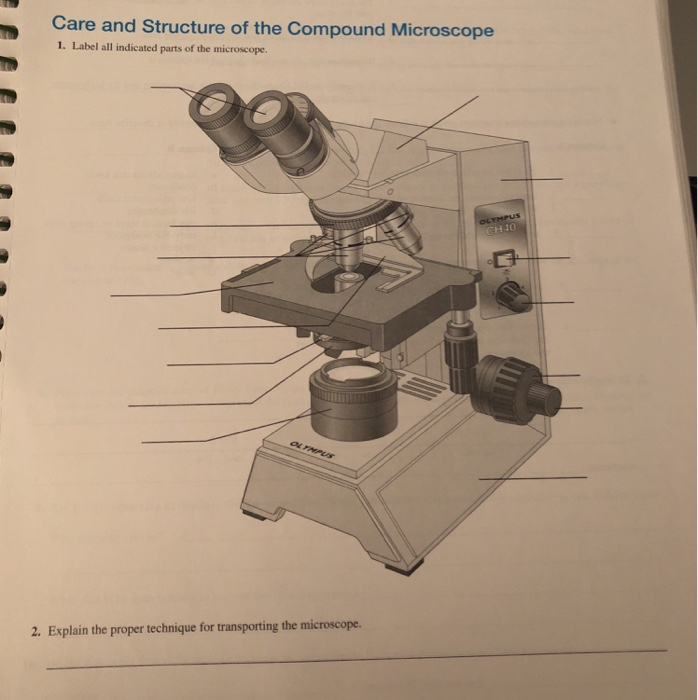
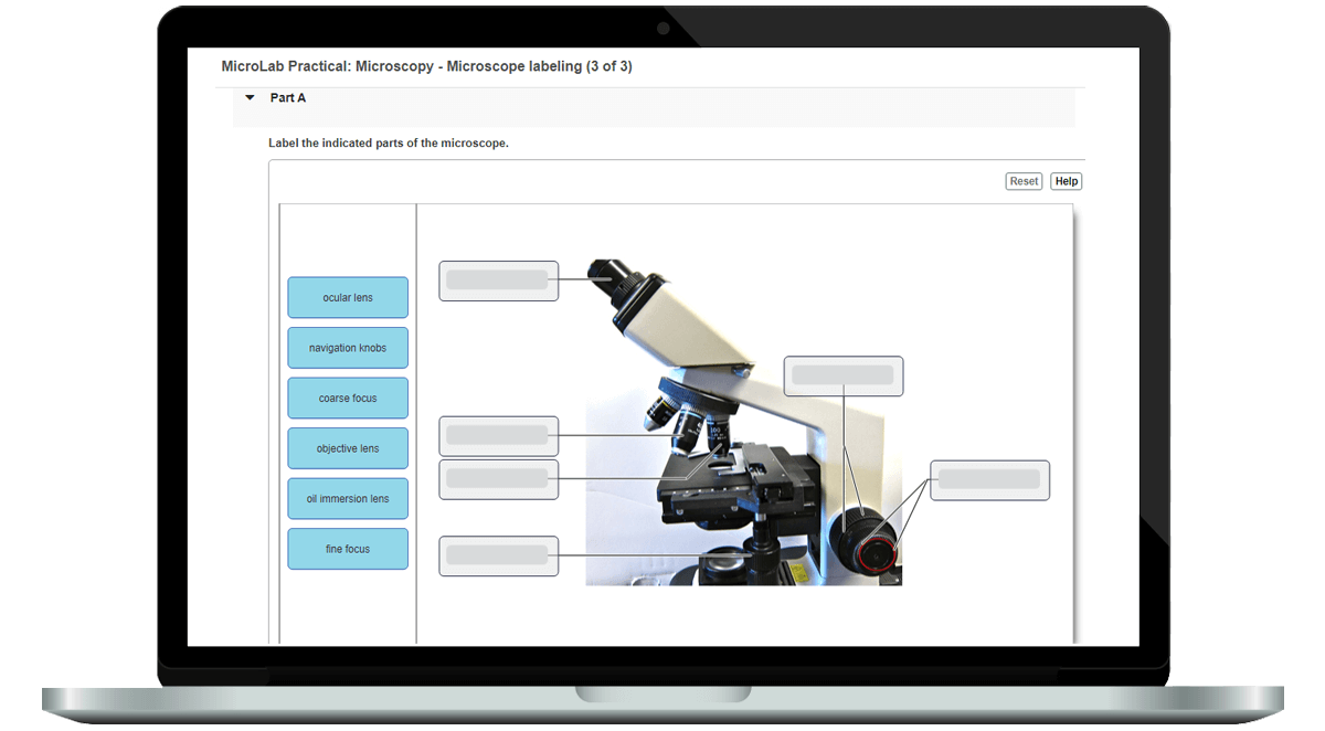
Label the indicated parts of the microscope
Microscope Parts and Functions Here are the important compound microscope parts... Eyepiece: The lens the viewer looks through to see the specimen. The eyepiece usually contains a 10X or 15X power lens. Diopter Adjustment: Useful as a means to change focus on one eyepiece so as to correct for any difference in vision between your two eyes. Parts of the Microscope with Labeling (also Free Printouts) A microscope is one of the invaluable tools in the laboratory setting. It is used to observe things that cannot be seen by the naked eye. Table of Contents 1. Eyepiece 2. Body tube/Head 3. Turret/Nose piece 4. Objective lenses 5. Knobs (fine and coarse) 6. Stage and stage clips 7. Aperture 9. Condenser 10. Condenser focus knob 11. Iris diaphragm 16 Parts of a Compound Microscope: Diagrams and Video The 16 core parts of a compound microscope are: Head (Body) Arm Base Eyepiece Eyepiece tube Objective lenses Revolving Nosepiece (Turret) Rack stop Coarse adjustment knobs Fine adjustment knobs Stage Stage clips Aperture Illuminator Condenser Diaphragm Video: Parts of a compound Microscope with Diagram Explained
Label the indicated parts of the microscope. MasteringMicrobiology - Week 1 Post Lab Flashcards | Quizlet Label the indicated parts of the microscope. (From top to bottom and left to right) Ocular Lens Objective Lens Oil Immersion Lens Navigation Knobs Coarse Focus Fine Focus Which of the following describes streptococci? Chains of spherically shaped cells Which of the following describes staphylococci? Clusters of spherically shaped cells Parts of the Microscope Flashcards | Quizlet Usually, a compound microscope has four objective lens: scanning (4x), low-power (10x), high-power (40x), and oil immersion (100x). Typical magnifying powers for the objectives are listed in parentheses Ocular lenses Binocular microscopes will have two lenses located in the eyepieces as the superior end of the head. compound microscope parts (labeling) Flashcards | Quizlet compound microscope parts (labeling) Term 1 / 14 eyepiece tube - connects the eyepiece to the objective lens Click the card to flip 👆 Definition 1 / 14 what is 1? Click the card to flip 👆 Flashcards Learn Test Match Created by barnettlily Terms in this set (14) eyepiece tube - connects the eyepiece to the objective lens what is 1? Parts of a Microscope - SmartSchool Systems Eyepiece lens magnifies the image of the specimen. This part is also known as ocular. Most school microscopes have an eyepiece with 10X magnification. 2. Eyepiece Tube or Body Tube. The tube hold the eyepiece. 3. Nosepiece. Nosepiece holds the objective lenses and is sometimes called a revolving turret.
List: Parts of a Microscope and their Function | Pathwooded Microscope Eyepiece or Ocular Lens. Located at the top of the microscope, the eyepiece is the lens assembly closest to your eye. It's the part of your microscope that you will look through to study objects and specimens. The eyepiece or ocular lens of a light microscope usually has a magnification level of 10x or 15x, but this can vary ... Label parts on microscope Flashcards | Quizlet Label parts on microscope. 4.0 (4 reviews) Term. 1 / 14. Eyepiece. Click the card to flip 👆. Definition. 1 / 14. Click the card to flip 👆. LABEL ALL INDICATED PARTS OF THE MICROSCOPE.docx - LABEL ALL INDICATED ... LABEL ALL INDICATED PARTS OF THE MICROSCOPE. 4 LABEL ALL INDICATED PARTS OF THE MICROSCOPE. 5 LABEL ALL INDICATED PARTS OF THE MICROSCOPE. 6 EXPLAIN THE PROPER TECHNIQUE FOR TRANSPORTING THE MICROSCOPE. WHEN TRANSPORTING THE MICROSCOPE, HOLD IT IN AN UPRIGHT POSITIONWITH ONE HAND ON ITS ARM AND THE OTHER SUPPORTING ITS BASE. Microscope: Parts Of A Microscope With Functions And Labeled Diagram. List down the 18 parts of a Microscope. Ocular Lens (Eye Piece) Diopter Adjustment Head Nose Piece Objective Lens Arm (Carrying Handle) Mechanical Stage Stage Clip Aperture Diaphragm Condenser Coarse Adjustment Fine Adjustment Illuminator (Light Source) Stage Controls Base Brightness Adjustment Light Switch
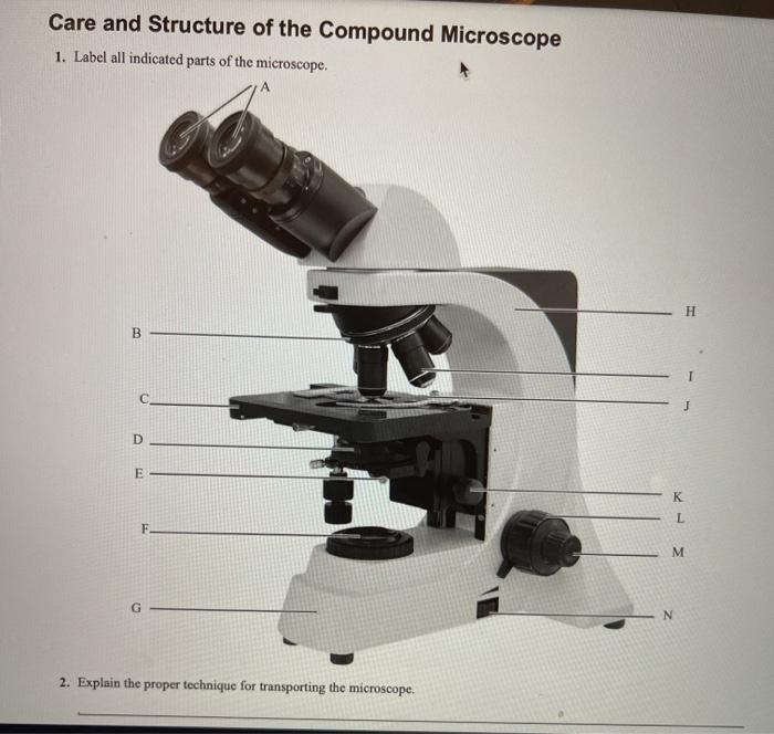
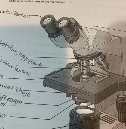
Mastering Microbiology Ch 3 Flashcards | Quizlet Drag the labels in the left column to indicate the microscope part that performs each function listed on the right. 1. Fine focus knob:used after initial focusing to sharpen the image 2. Ocular lens:lens that you look through 3. Objective lens:lens that is closest to the slide and provides initial magnification of a specimen 4. Microscope Parts & Functions - AmScope Head: The upper part of the microscope houses the eyepiece and objective lenses. Tube: Where the eyepieces are dropped in.Also, it connects the eyepieces to the objective lenses. Stage: The flat platform that supports the slides.Stage clips hold the slides in place. If your microscope has a mechanical stage, the slide is controlled by turning two knobs instead of having to move it manually. PDF The Microscope - Holly H. Nash-Rule, PhD After the parts of the microscope have been identified, turn on the light and adjust the interpupillary distance so that ... Care and Structure of the Compound Microscope 1. Label all indicated parts of the microscope. Ocular lenses Rotating nosepiece Objective lenses Stage Mechanical stage Iris diaphragm lever Condenser Substage light Head Arm 16 Parts of a Compound Microscope: Diagrams and Video The 16 core parts of a compound microscope are: Head (Body) Arm Base Eyepiece Eyepiece tube Objective lenses Revolving Nosepiece (Turret) Rack stop Coarse adjustment knobs Fine adjustment knobs Stage Stage clips Aperture Illuminator Condenser Diaphragm Video: Parts of a compound Microscope with Diagram Explained
Solved Care and Structure of the Compound Microscope 1 ...
Parts of the Microscope with Labeling (also Free Printouts) A microscope is one of the invaluable tools in the laboratory setting. It is used to observe things that cannot be seen by the naked eye. Table of Contents 1. Eyepiece 2. Body tube/Head 3. Turret/Nose piece 4. Objective lenses 5. Knobs (fine and coarse) 6. Stage and stage clips 7. Aperture 9. Condenser 10. Condenser focus knob 11. Iris diaphragm
Solved Care and Structure of the Compound Microscope 1 ...
Microscope Parts and Functions Here are the important compound microscope parts... Eyepiece: The lens the viewer looks through to see the specimen. The eyepiece usually contains a 10X or 15X power lens. Diopter Adjustment: Useful as a means to change focus on one eyepiece so as to correct for any difference in vision between your two eyes.
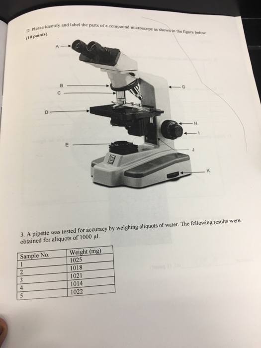
Solved Identify and label the parts of a compound microscope ...

Microscope Parts & Functions - AmScope
Microscope Parts Quiz

Parts of Stereo Microscope (Dissecting microscope) – labeled ...

Label the Microscope | Microscope parts, Teaching biology ...

MCB2004L Microscopy Flashcards | Quizlet
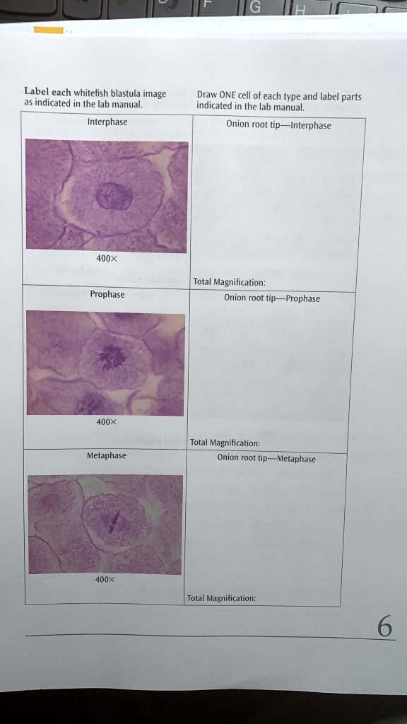
SOLVED: Label each whitefish blastula image indicated in the ...
Solved Care and Structure of the Compound Microscope 1 ...

Label the indicated structures in this diagram for eukaryotic ...

Microscope Labeling Diagram | Quizlet

Microscope Diagram Labeled, Unlabeled and Blank | Parts of a ...
Solved Care and Structure of the Compound Microscope 1 ...

Modular low-light microscope for imaging cellular ...
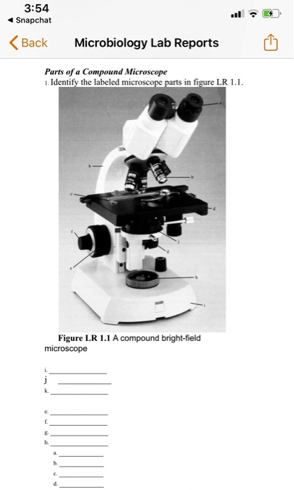
SOLVED: 3:54 Snapchat Back Microbiology Lab Reports Parts of ...
Microscope Quiz
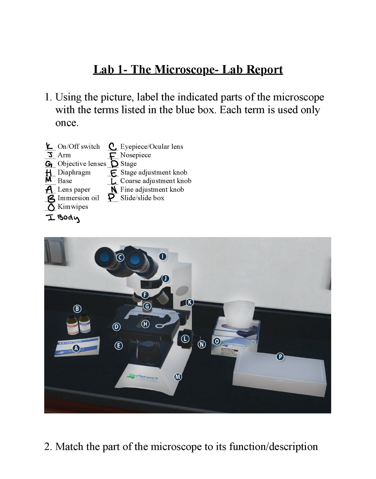
Lab 1- The Microscope- Lab Report 3 - Using the picture ...

Parts of a Microscope Diagram | Quizlet

The Microscope
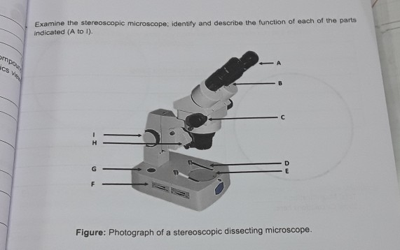
Solved Examine the stereoscopic microscope; identify and ...
Parts Of The Microscope Label Teaching Resources | TPT
Solved) - Care and Structure of the Compound Microscope 1 ...
Parts of a Microscope Quiz
Activity 1 Review of Microscopy
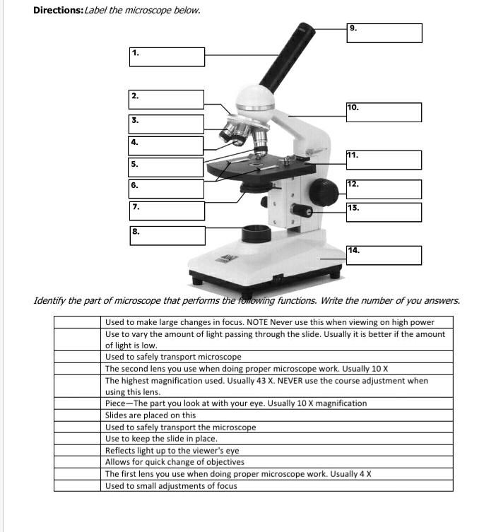
Solved Directions:Label the microscope below. 9. 1. 111 2 ...
REVIEW SHEET The Microscope lopMLA Volahio noeaes adoaSe

Lab Chapter #2 - The Microscope Diagram | Quizlet

Amazon.com: Upgrade Semi-Automatic Sheet Embosser Metal ...

This is a common compound microscope. Label its parts from A ...

Axodendritic synapses, TEM - Stock Image - C053/4784 ...

In Silico Labeling: Predicting Fluorescent Labels in ...
Parts of the Microscope with Labeling (also Free Printouts ...

Compound Microscope Parts – Labeled Diagram and their ...

Microplastics and Nanoplastics: Emerging Contaminants in Food ...
bio exercise 3&4 fahmida Usman

Basic parts of a circular saw machine (29). | Download ...
Features | Educators | Mastering Microbiology | Pearson

Parts of a microscope with functions and labeled diagram
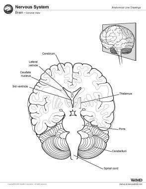
Ventricles of the Brain: Overview, Gross Anatomy, Microscopic ...
Komentar
Posting Komentar