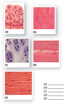
39 label the photomicrograph
Final Exam A&P 1 Flashcards | Quizlet Label the structures of the skin and subcutaneous tissue. Label the photomicrograph of thick skin. Epidermis, stratum corneum, stratum lucidum, stratum granulosum, stratum spinosum, stratum basale, dermis Ch 21 assignment 2 Flashcards | Quizlet Correctly label the anatomical features of lymphatic capillaries. Which of the following is a function of the lymphatic system? Check all that apply. Recover fluid from the interstium to the blood plasma. Remove foreign matter from fluid before returning it to the bloodstream. Absorb dietary lipids. Label the photomicrograph based on the hints ...
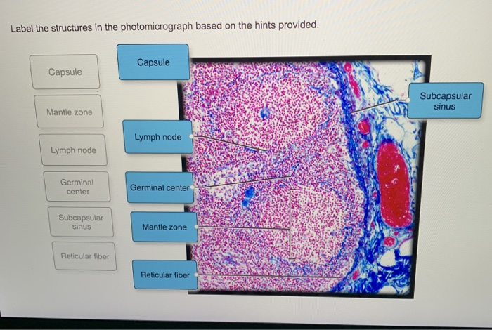
Ch. 22 Assessment Flashcards | Quizlet Study with Quizlet and memorize flashcards containing terms like Label the photomicrograph based on the hints provided., Label the photomicrograph based on the hints provided., 1. Foreign antigen description: 2. Foreign antigen example: 3. Self-antigen description: 4. Self-antigen example: and more.

Label the photomicrograph
anatomy lab, exam 3, lab 9, Spinal Nerves, Integument, and ... in general, nerves from the posterior division of the brachial plexus tend to innervate muscles that extend the parts of the upper limb. Name the highlighted nerve that provides some of the innervation to joints of the hands. Label the structures of the skin and subcutaneous tissues. Match the label to its appropriate spinal cord component. BIOL 320 - Practical 2 - Lab 7 Flashcards | Quizlet Label the types of cells in the photomicrograph using the hints provided. Label the photomicrograph of the wall of the aorta using the hints provided. Label the photomicrograph of the wall of the aorta using the hints provided. Label the photomicrograph of the wall of the inferior vena cava using the hints provided. tunica intima of arteriole. Lab 1 Homework BIOL 320 Flashcards | Quizlet Label the photomicrograph of the wall of the aorta using the hints provided. Correctly label the parts of centrifuged blood. Match each phrase to the formed element it describes.
Label the photomicrograph. Solved Label the photomicrograph. Myoepithelial cell Lumen ... Expert Answer. 94% (16 ratings) The given micrograph is labelled and is attached below: Justification: Epithelial cell: The epithelial cells in mammary …. View the full answer. Transcribed image text: Label the photomicrograph. Myoepithelial cell Lumen Epithelial cell Apocrine sweat gland OMG Den Stree. Previous question Next question. Lab 1 Homework BIOL 320 Flashcards | Quizlet Label the photomicrograph of the wall of the aorta using the hints provided. Correctly label the parts of centrifuged blood. Match each phrase to the formed element it describes. BIOL 320 - Practical 2 - Lab 7 Flashcards | Quizlet Label the types of cells in the photomicrograph using the hints provided. Label the photomicrograph of the wall of the aorta using the hints provided. Label the photomicrograph of the wall of the aorta using the hints provided. Label the photomicrograph of the wall of the inferior vena cava using the hints provided. tunica intima of arteriole. anatomy lab, exam 3, lab 9, Spinal Nerves, Integument, and ... in general, nerves from the posterior division of the brachial plexus tend to innervate muscles that extend the parts of the upper limb. Name the highlighted nerve that provides some of the innervation to joints of the hands. Label the structures of the skin and subcutaneous tissues. Match the label to its appropriate spinal cord component.
Solved Label the photomicrograph of thin skin | Chegg.com

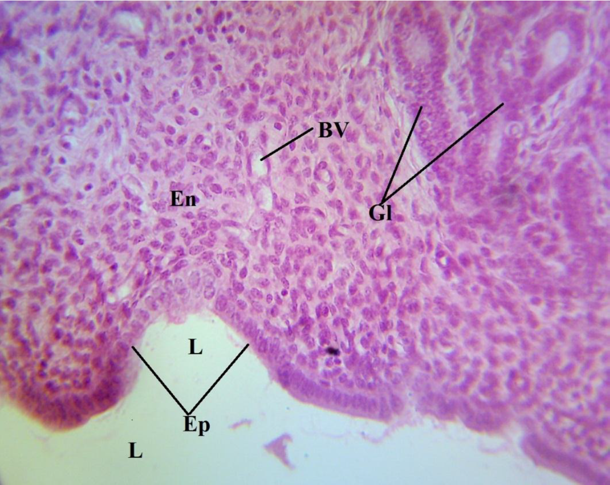
Solved Label the photomicrograph based on the hints | Chegg.com
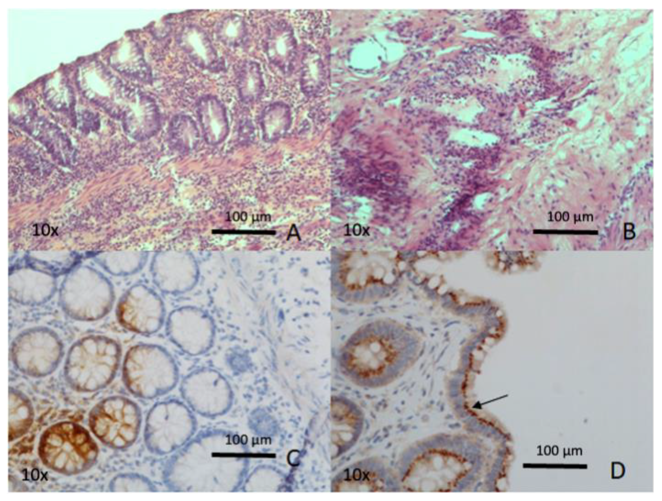
Biomedicines | Free Full-Text | Intestinal Ischemia: Unusual ...
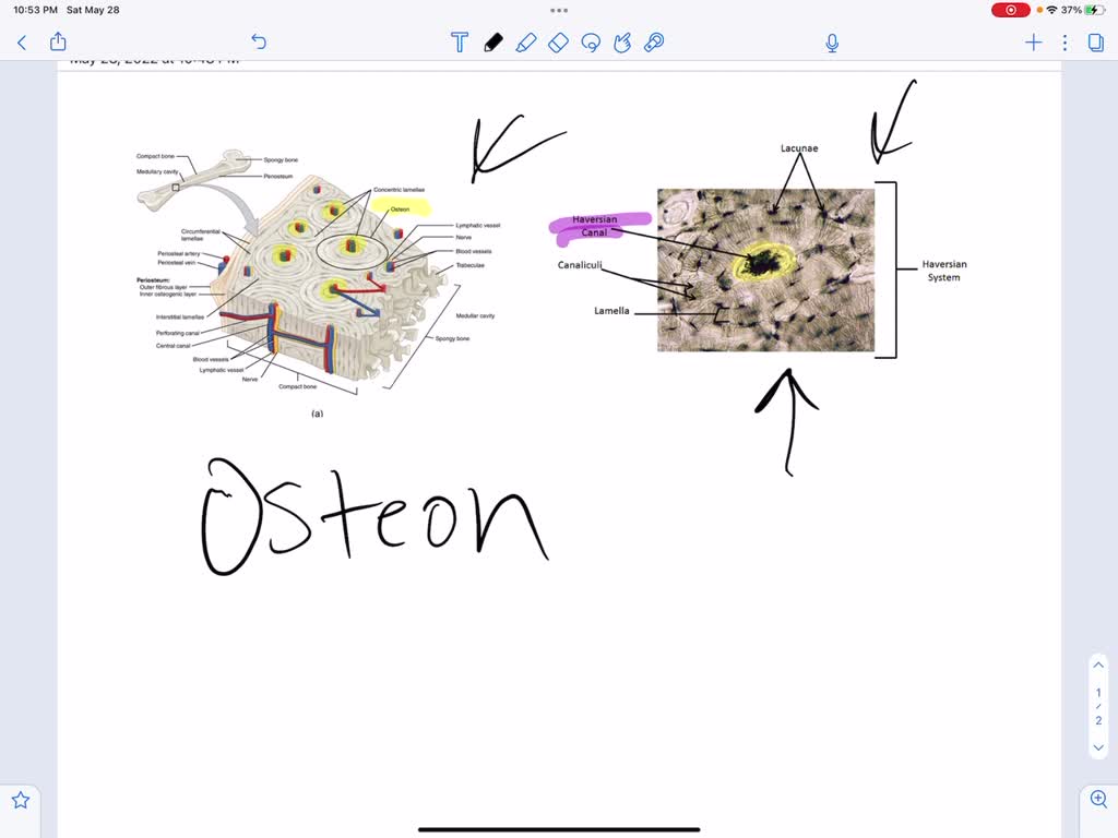
"In the photomicrograph below of compact bone tissue, find and label the indicated structures, Osleon, Lamella, Lacuna, Osteocyte, Canallcull, Central canal, Obtain a slide of ground compact bone ...
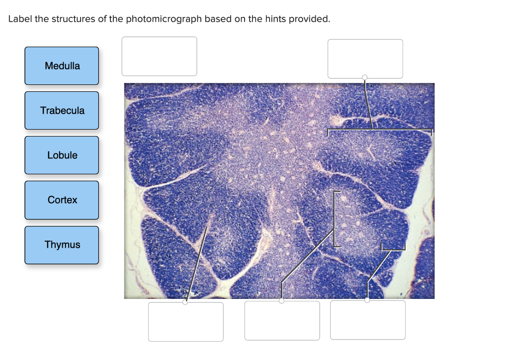
Solved Label the structures of the photomicrograph based on ...

Tamu Biol 320: Module 7 Flashcards | Quizlet
9700 QR Dynamic Papers Biology al Cambridge
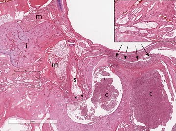
Syed Salahuddin Ahmed on Twitter: "#image #histopath #bone ...
Brm Inhibits the Proliferative Response of Keratinocytes and ...

Human blood smear under microscope, light photomicrograph ...

Pin by nico x. on Anatomy | Games, Tetris, Anatomy
Solved Label the structures in the photomicrograph based on ...
Photomicrograph depicting Histomonas meleagridis - Texas A&M ...

Endocrine Lab Flashcards | Quizlet
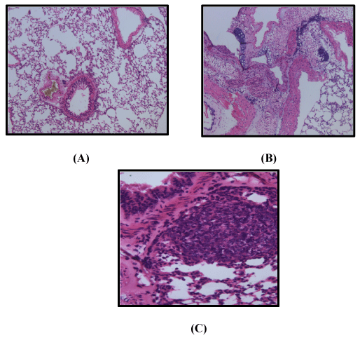
photomicrograph of lung sections

Photomicrograph of Thin Skin Quiz

View Image

Photomicrograph Liver Biopsy Histology Pathology Showing ...
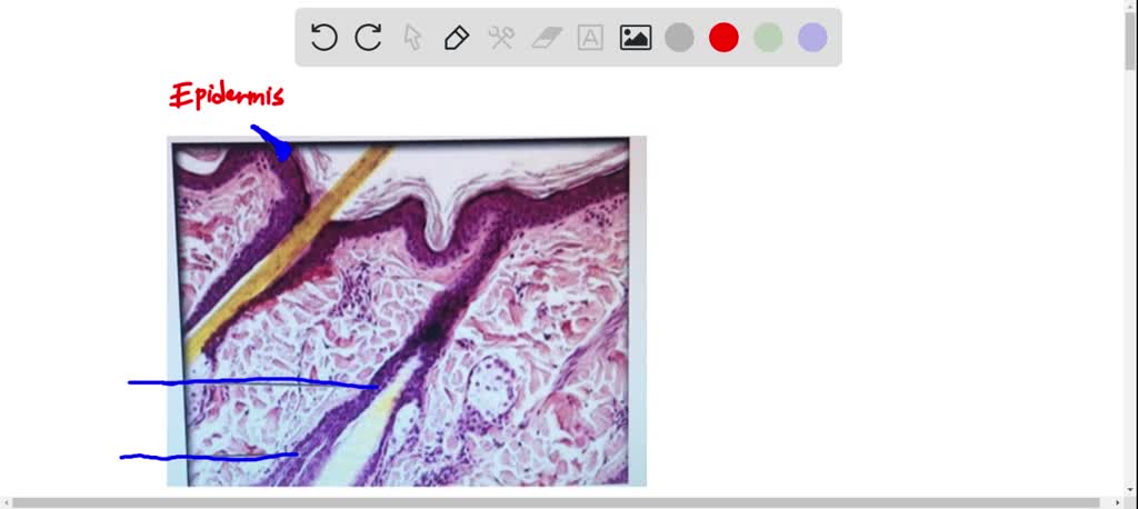
Label tne photomicrograph Of the Skin and Its accessory structures, Sebaceous gland, Duct ofl, sebaceous gland, Epidermis, Hair follicle

APR Lymphatic System Flashcards | Quizlet
Animals | Free Full-Text | The Formation and Invariance of ...

Solved Endocrine Lab Worksheet Label the photomicrograph ...
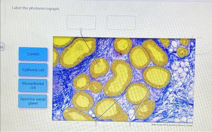
Solved Label the photomicrograph. Lumen Epithelial cell ...

Light photomicrograph of Cucurbita stem cross section seen ...
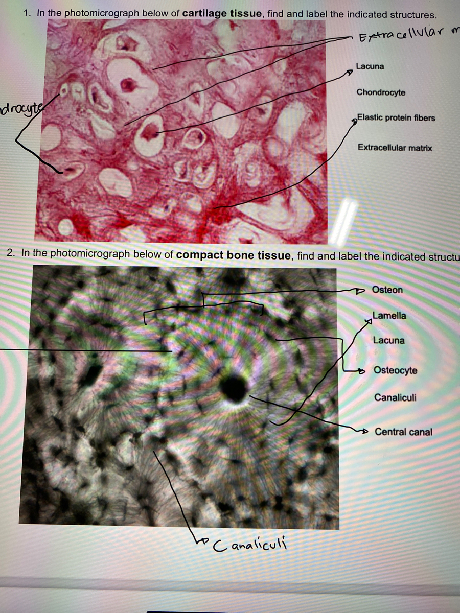
Answered: 1. In the photomicrograph below of… | bartleby

A & P lab test 4 Flashcards | Quizlet
Bilateral tympanokeratomas (cholesteatomas) with bilateral ...

Ch. 22 Assessment Flashcards | Quizlet
Get Answer) - Label the following photomicrographs with the ...

Human scalp, light micrograph - Stock Image - C054/3494 ...
Effects of ethanol extracts of Diodia sarmentosa leaves on ...

View Image

Cartilage in fetal finger. Photomicrograph Stock Photo - Alamy

BIOL 320 - Practical 2 - Lab 7 Flashcards | Quizlet

View Image

Label the photomicrograph based on the hints provided ...

Bio lab - Type your answers in a comment, the first three ...

Page 2 | Metastasis Images - Free Download on Freepik
Solved] Lab 3 Exercise 3-4 LAB 3 EXERCISE 3-4 1. In the ...


Komentar
Posting Komentar