38 microscope labeled diagram
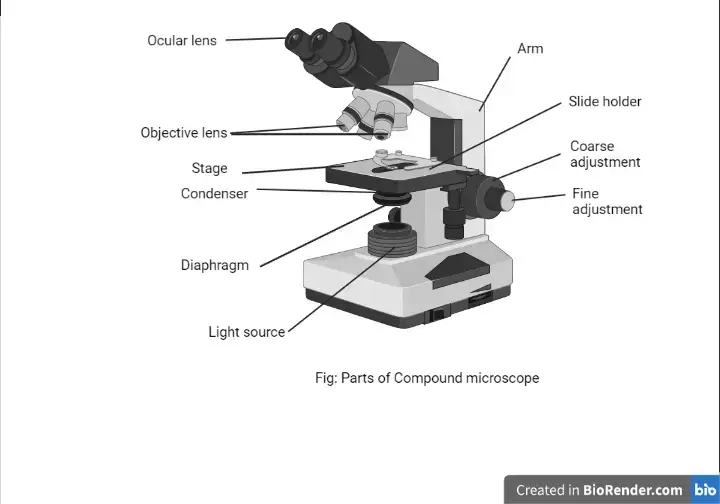
microscopeinternational.com › compound-microscopeCompound Microscope Parts, Functions, and Labeled Diagram Nov 18, 2020 · Base: Bottom base of the microscope that houses the illumination & supports the compound microscope. Objective lenses: There are usually 3-5 optical lens objectives on a compound microscope each with different magnification levels. 4x, 10x, 40x, and 100x are the most common magnifying powers used for the objectives. anatomylearner.com › simple-squamous-epitheliumSimple Squamous Epithelium under a Microscope with a Labeled ... Mar 4, 2022 · Here the artery labeled diagram shows the tunica intima that consists of endothelium, basal lamina, subendothelium connective tissue, and internal elastic lamina. You will find the endoplasmic reticulum and mitochondria in the cytoplasm of the endothelium cell under the electron microscope.
microbenotes.com › electron-microscope-principleElectron Microscope- Definition, Principle, Types, Uses ... Apr 4, 2022 · An electron microscope is a microscope that uses a beam of accelerated electrons as a source of illumination. It is a special type of microscope having a high resolution of images, able to magnify objects in nanometres, which are formed by controlled use of electrons in a vacuum captured on a phosphorescent screen.

Microscope labeled diagram
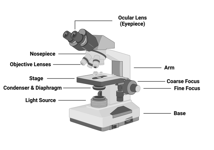
microscopewiki.com › compound-microscopeCompound Microscope – Diagram (Parts labelled), Principle and ... Oct 10, 2022 · See: Labeled Diagram showing differences between compound and simple microscope parts. Structural Components. The three structural components include. 1. Head. This is the upper part of the microscope that houses the optical parts. 2. Arm . This part connects the head with the base and provides stability to the microscope. microbenotes.com › parts-of-a-microscopeParts of a microscope with functions and labeled diagram Sep 17, 2022 · Light Microscope- Definition, Principle, Types, Parts, Labeled Diagram, Magnification Amazing 27 Things Under The Microscope With Diagrams Plant Cell- Definition, Structure, Parts, Functions, Labeled Diagram rsscience.com › compound-microscope-parts-labeledCompound Microscope Parts – Labeled Diagram and their ... There are three major structural parts of a microscope: Head, Base, and Arm. Always lift a microscope by holding both the arm and base with two hands. There are two major optical lens parts of a microscope: Eyepiece (10x) and Objective lenses (4x, 10x, 40x, 100x).
Microscope labeled diagram. microbenotes.com › compound-microscope-principleCompound Microscope- Definition, Labeled Diagram, Principle ... Apr 3, 2022 · Light Microscope- Definition, Principle, Types, Parts, Labeled Diagram, Magnification Amazing 27 Things Under The Microscope With Diagrams 22 Types of Spectroscopy with Definition, Principle, Steps, Uses rsscience.com › compound-microscope-parts-labeledCompound Microscope Parts – Labeled Diagram and their ... There are three major structural parts of a microscope: Head, Base, and Arm. Always lift a microscope by holding both the arm and base with two hands. There are two major optical lens parts of a microscope: Eyepiece (10x) and Objective lenses (4x, 10x, 40x, 100x). microbenotes.com › parts-of-a-microscopeParts of a microscope with functions and labeled diagram Sep 17, 2022 · Light Microscope- Definition, Principle, Types, Parts, Labeled Diagram, Magnification Amazing 27 Things Under The Microscope With Diagrams Plant Cell- Definition, Structure, Parts, Functions, Labeled Diagram microscopewiki.com › compound-microscopeCompound Microscope – Diagram (Parts labelled), Principle and ... Oct 10, 2022 · See: Labeled Diagram showing differences between compound and simple microscope parts. Structural Components. The three structural components include. 1. Head. This is the upper part of the microscope that houses the optical parts. 2. Arm . This part connects the head with the base and provides stability to the microscope.
Microscope Diagram and Functions | Microscope parts, Science ...
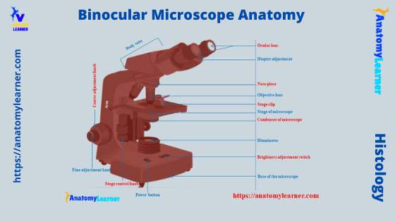
Binocular Microscope Anatomy - Parts and Functions with a ...

Solved A. OLYMPUS C. B. Use the Diagram to answer the | Chegg.com

Label The Microscope Parts! Diagram | Quizlet

Different types of Microscopes – light microscope, electron ...

Compound Microscope Parts – Labeled Diagram and their ...
Compound Microscope Parts, Functions, and Labeled Diagram ...
Addgene: Using a Light Microscope Protocol

Microscope Labeling Part 1 Diagram | Quizlet

Draw a well labelled diagram of a microscope. - Brainly.in

Microscope Diagram Labeled, Unlabeled and Blank | Parts of a ...
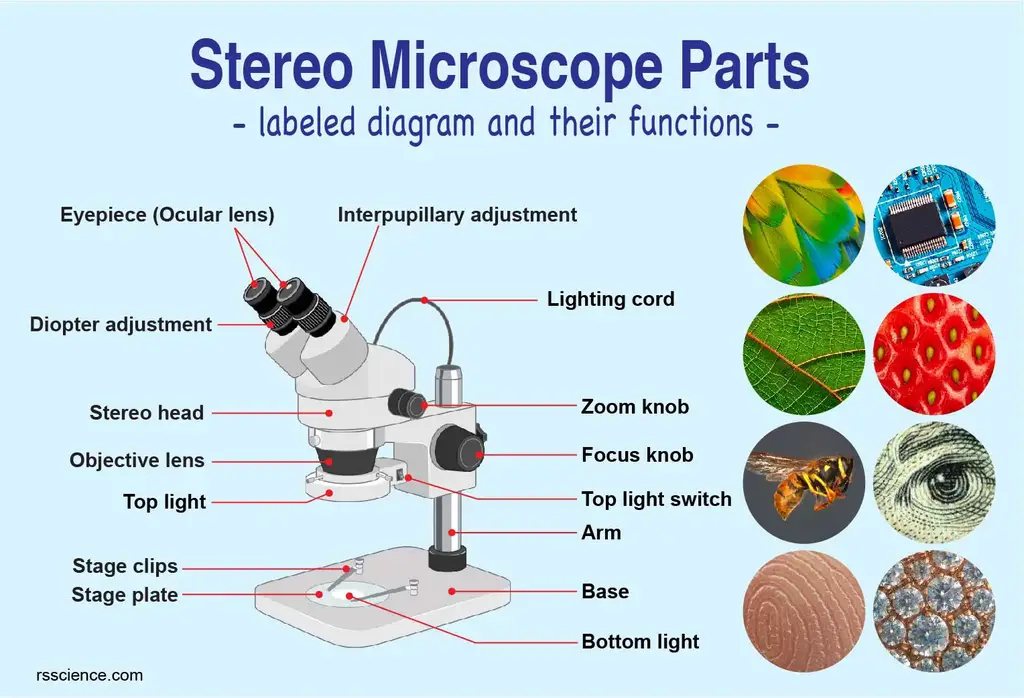
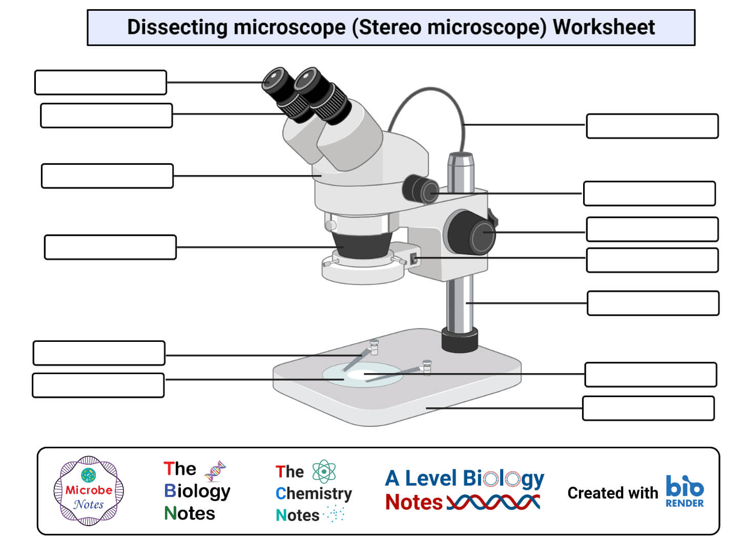
Parts of Stereo Microscope (Dissecting microscope) – labeled ...

Label the Microscope Diagram | Download Scientific Diagram

Labelling a Microscope Diagram | Quizlet
Parts of a microscope with functions and labeled diagram
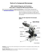
MICROBIO 16 Parts of a Compound Microscope with Diagram and ...

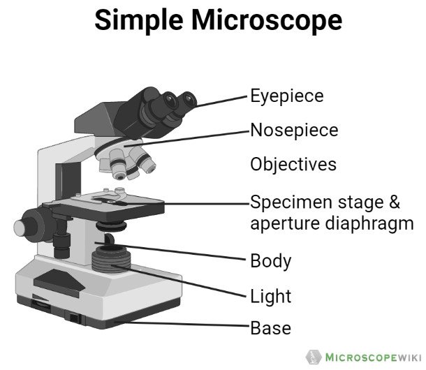
Simple Microscope - Diagram (Parts labelled), Principle ...

This is a common compound microscope. Label its parts from A ...
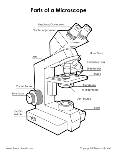
Microscope Diagram - Tim's Printables

Biology Lab Microscope Labeling Diagram | Quizlet

Living Environment Course
Parts of a Microscope Foldable-Labeled by Sciencerly | TPT
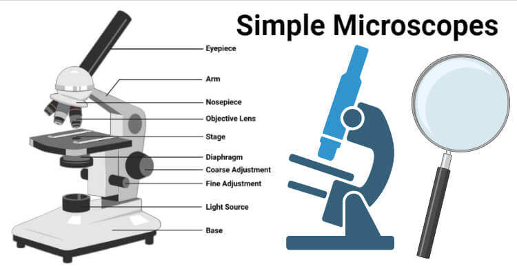
Simple Microscope- Definition, Principle, Magnification ...

Compound Microscope Parts – Labeled Diagram and their ...
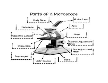
Parts of the Microscope (Labeled Diagrams) - Simple and ...

Diagram of a Compound Microscope

label microscope diagram | Charts | Microscope, Diagram chart ...

Labeling the Parts of the Microscope | Microscope activity ...

Light Microscope- Definition, Principle, Types, Parts ...
Label a microscope - Teaching resources

Microscope Labeling Diagram | Quizlet
Diagram of a Microscope - Guide to using a microscope
Microscope Types (with labeled diagrams) and Functions

Microscope With Labels clip art | Microscope parts, Science ...

Lable the microscope worksheet
Parts of a Microscope with Their Functions – Microbe Online
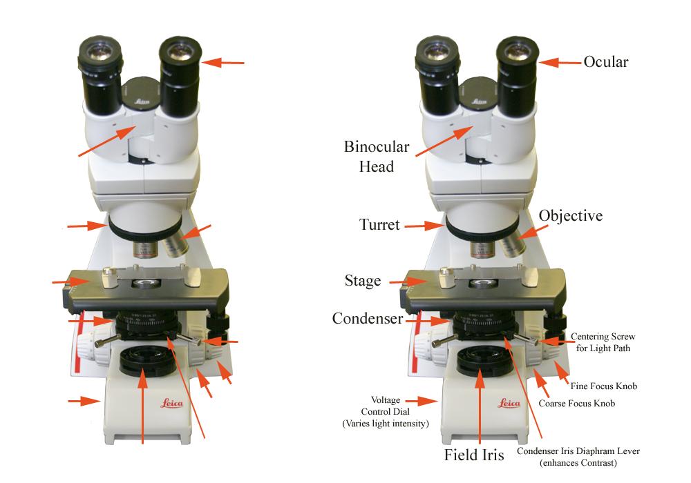
Front view of the Leica microscope used in General Botany ...

File:Labelledmicroscope.gif - Wikimedia Commons


Komentar
Posting Komentar