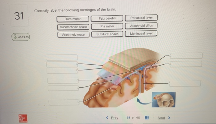
45 correctly label the following meninges of the brain.
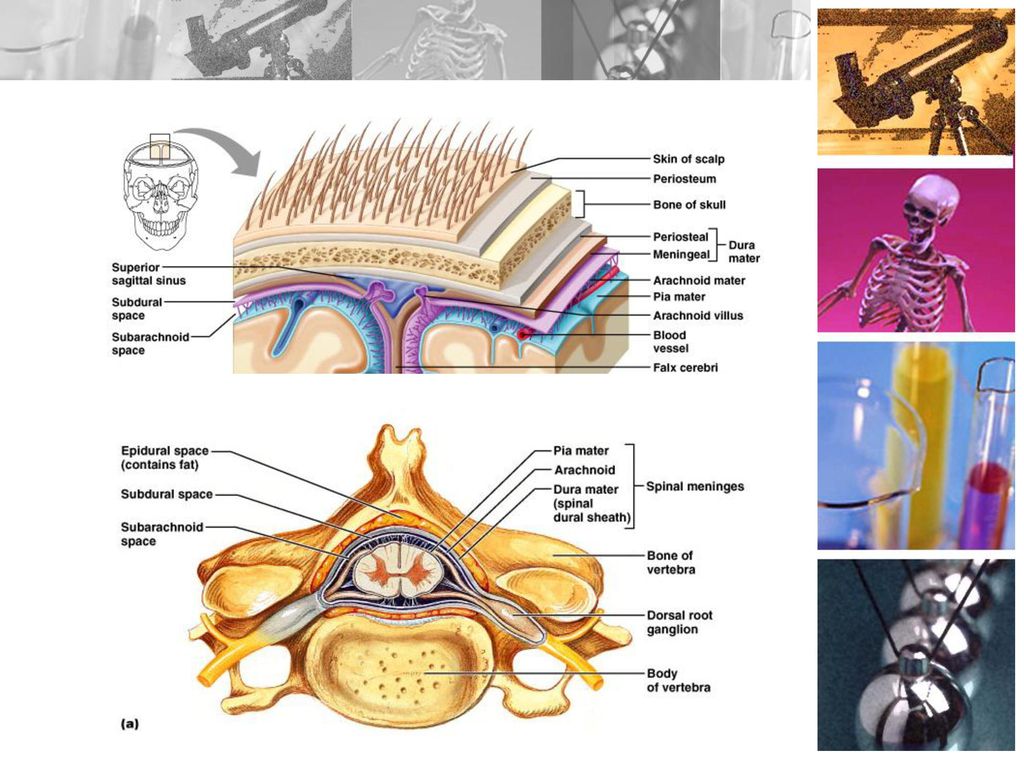
PDF The Brain - Overview - Vermont __T__ 6. Neurons are the basic brain cells. __T _ 7. The brain communicates via chemicals. _F __ 8. Neurotransmitters pass directly from the axon. to the dendrite. MAY PASS AXON TO AXON, AXON TO DENDRITE AND DENDRITE TO DENDRITE. __T__ 9. The brain continues to grow new neurons after. birth. __T_ 10. Each lobe of the brain has specific ... PDF Brain Anatomy - Wou BI 335 - Advanced Human Anatomy and Physiology Western Oregon University Figure 4: Mid-sagittal section of brain showing diencephalon (includes corpus callosum, fornix, and anterior commissure) Marieb & Hoehn (Human Anatomy and Physiology, 9th ed.) - Figure 12.10 Exercise 2: Utilize the model of the human brain to locate the following structures / landmarks for the
Draw a labelled diagram of human brain and write its any two functions. Forebrain: It has the following parts: 1. Cerebrum: Which performs thinking, reasoning, speech, intelligence and usage of information. 2. Olfactory: Where lobes are responsible for detecting the smell from different receptors. 3. Diencephalon: Which controls body temperature, the urge of eating, drinking, etc. Was this answer helpful? 0.

Correctly label the following meninges of the brain.
Positions and Functions of the Four Brain Lobes | MD-Health.com The occipital lobe, the smallest of the four lobes of the brain, is located near the posterior region of the cerebral cortex, near the back of the skull. The occipital lobe is the primary visual processing center of the brain. Here are some other functions of the occipital lobe: Visual-spatial processing. Movement and color recognition. 5 Lobes Of The Brain (A Complete Guide) - NeuroTray 5 lobes of the brain. Each cerebral hemisphere is divided into five lobes: the frontal lobe, the parietal lobe, the occipital lobe and the temporal lobe, four of which have the same name as the bone above them. Deep within the lateral sulcus lies a fifth lobe, the insula or Island of Reil. PDF Brain Review and Wkst Answer - Mayfield City Schools Created Date: 4/30/2013 4:05:46 PM
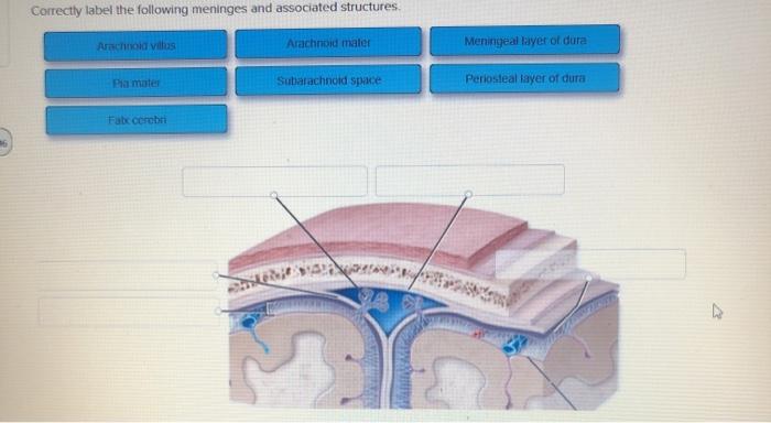
Correctly label the following meninges of the brain.. Correctly label the following meninges of the brain....open 8 Correctly label the following meninges of the brain. Arachnoid villus Arachnoid mater Subdural space Meningeal layer Pia mater Periosteal layer Dura mater: Subarachnoid space Falx cerebri Dura mater: Reset Zoom Human Brain - Structure, Diagram, Parts Of Human Brain - BYJUS The human brain controls nearly every aspect of the human body ranging from physiological functions to cognitive abilities. It functions by receiving and sending signals via neurons to different parts of the body. The human brain, just like most other mammals, has the same basic structure, but it is better developed than any other mammalian brain. Spinal Meninges Anatomy, Diagram & Function | Body Maps - Healthline Pia mater: The innermost layer, the pia mater hugs the spinal cord and brain like a coat. It has blood vessels that deliver oxygen and nutrients to the spinal cord. To check for problems of the ... Study Guide.docx - Correctly match each nerve ending with... Correctly identify the following anatomical landmarks for the olfactory projection pathways in the brain. Correctly label the following anatomical features of the eye. Drag each label to the appropriate box to indicate whether each statement is associated with rods or cones.
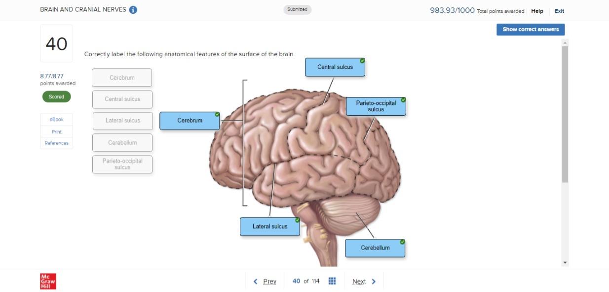
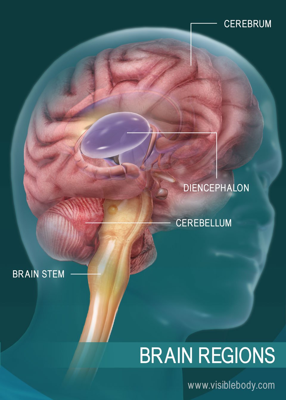
Brain labeling quizlet - wclclm.allwallets.shop brain, the mass of nerve tissue in the anterior end of an organism. The brain integrates sensory information and directs motor responses; in higher vertebrates it is also the centre of learning. The human brain weighs approximately 1.4 kg (3 pounds) and is made up of billions of cells called neurons. Junctions between neurons, known as synapses ... Diagram of the Brain and its Functions - Bodytomy Functions. The frontal lobe is involved with the main executive functions of the brain, which include: Judgment, that is, the ability to recognize future consequences resulting from ongoing actions. This activity mostly occurs in the pre-frontal area. Analytical and critical reasoning. Chapter 14 Worksheet Flashcards | Quizlet Study with Quizlet and memorize flashcards containing terms like Correctly label the following anatomical features of the surface of the brain., Correctly label the following meninges of the brain., Place a single word into each sentence to make it correct, then place each sentence into a logical paragraph order describing the flow of cerebrospinal fluid. and more. Brain Structure And Function | Brain Injury | British Columbia The Cerebellum - The cerebellum, or "little brain", is similar to the cerebrum with its two hemispheres and highly folded surface. It is associated with regulation and coordination of movement, posture, balance and cardiac, respiratory and vasomotor centers. Brain Stem - The brain stem is located beneath the limbic system.
The Ventricles of the Brain - Lateral - Third - TeachMeAnatomy The ventricular system is a set of communicating cavities within the brain. These structures are responsible for the production, transport and removal of cerebrospinal fluid, which bathes the central nervous system. In this article, we shall look at the functions and production of cerebrospinal fluid, and the anatomy of the ventricles that contains it. (Get Answer) - The Thickest Of The Meninges Is The A. Pia Mater. B ... The Thickest Of The Meninges Is The A. Pia Mater. B Arachnoid Mater C. Subdural Space. D Subarachnoid Space. E. Dura Mater. Structurally, The Simplest Reflex Is The A. Stretch Reflex. B. Golgi Tendon Reflex. What midbrain structure is a visual reflex center Superior colliculi of the tectal plate The Meninges of the Brain Correctly label the following meninges and associated structures. The Flow of Cerebrospinal Fluid Place a single word into each sentence to make it correct. Nervous System - Label the Brain - TheInspiredInstructor.com Choose the correct names for the parts of the brain. ( 9) This brain part controls thinking. (10) This brain part controls balance, movement, and coordination. (11) This brain part controls involuntary actions such as breathing, heartbeats, and digestion. (12) This part of the nervous system moves messages between the brain and the body.
Synapse - Wikipedia
Solved Correctly label the following meninges of the brain. - Chegg Correctly label the following meninges of the brain. Arachnoid villus Arachnoid mater Subdural space Meningeal layer Pia mater Periosteal layer Dura mater: Subarachnoid space Falx cerebri Dura mater: Reset Zoom ; Question: Correctly label the following meninges of the brain. Arachnoid villus Arachnoid mater Subdural space Meningeal layer Pia ...
BRAIN AND CRANIAL NERVES BSC 2085 Flashcards | Chegg.com
A Labelled Diagram Of Neuron with Detailed Explanations - BYJUS Diagram Of Neuron. A neuron is a specialized cell, primarily involved in transmitting information through electrical and chemical signals. They are found in the brain, spinal cord and the peripheral nerves. A neuron is also known as the nerve cell. The structure of a neuron varies with their shape and size and it mainly depends upon their ...

CBIO Figures Flashcards | Quizlet
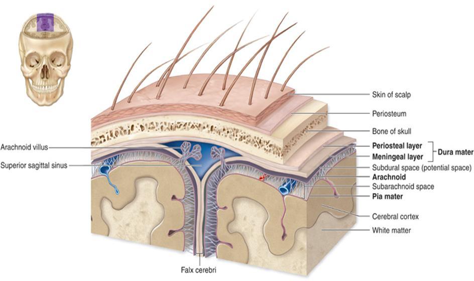
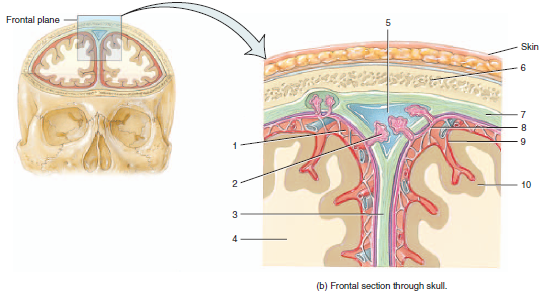
Meninges Layers, Function & Anatomy - Study.com The meninges are three fibrous membranes that enclose the central nervous system, which consists of the brain and spinal cord. The thick and tough outermost meningeal layer is called the dura mater .
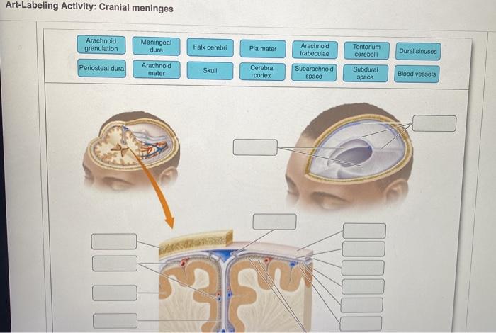
Solved Art-Labeling Activity: Cranial meninges Arachnold ...
Correctly Label the Following Anatomical Features of a Neuron. Correctly label the following anatomical features of the spinal cord. Free nerve endings subcutaneous layer oil gland dermis epidermis sweat gland sensory receptor adipose tissue sweat pore nerve. Label the nerves of the brachial plexus.

Meninges Layers, Function & Anatomy | Dura Mater, Arachnoid ...
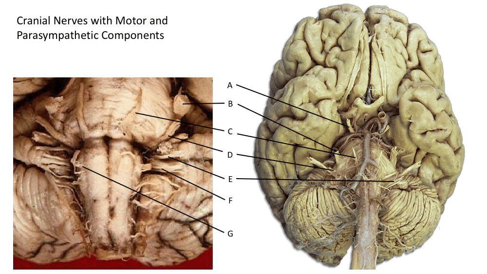
Brain & CN Worksheet Flashcards | Quizlet Consider a situation where a stroke or mechanical trauma has occurred resulting in damage to one of the areas of the brain indicated in the image. Drag each label into the proper location in order to identify the area that would most likely have been affected. Drag each of the given signs and symptoms of nerve damage to the proper position to ...

Match each of the meninges of the brain with the correct ...
Brain Regions and Their Functions - Bodytomy This article deals with the brain regions and their functions which will help you understand what part of your brain controls what mental activity. The nervous system comprises of the brain, spinal cord and sensory nerves and it is the most complicated human body system. The Brain is the most complex and delicate organ of the human body.
Solved Correctly label the following meninges of the brain ...
Meninges. Human brain | Human brain, Dura mater, Human anatomy and ... Oct 10, 2018 - Meninges. Human brain --- ZIP includes: - 1 files EPS8 - 1 files AI - 1 GPEG 4700x3000
Neuro Anatomy Flashcards | Chegg.com
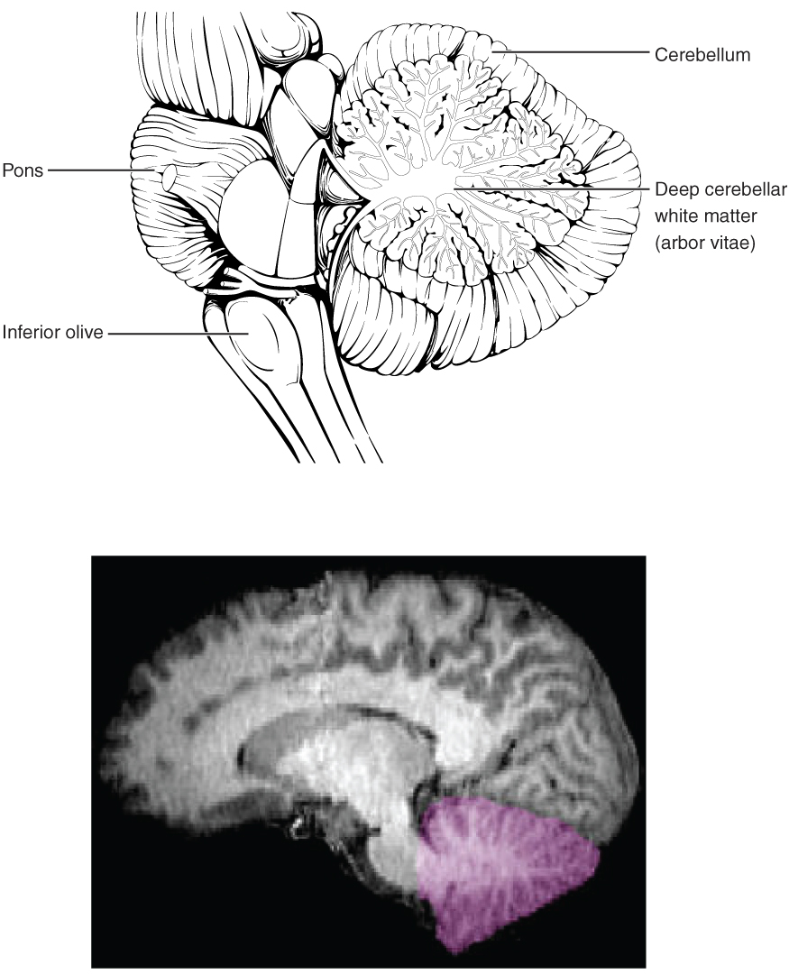
Brain: Anatomy, Pictures, Functions, and Conditions - Verywell Mind The brainstem is an area located at the base of the brain that contains structures vital for involuntary functions such as the heartbeat and breathing. The brain stem is comprised of the midbrain, pons, and medulla. Midbrain . The midbrain is often considered the smallest region of the brain. It acts as a sort of relay station for auditory and ...

Brain meninges labeled Diagram | Quizlet
From outermost to innermost, what are the names and the correct order ... Dura mater Arachnoid mater Pia mater From outermost to innermost, Meninges around the brain has three layers : 1. The dura mater 2. the arachnoid mater and 3. the pia mater. The dura mater has two layers : periosteal and meningeal. There is a space between the dura mater and the arachnoid mater, called subdural space. There is also a space between the arachnoid and the pia mater, which is ...
Frontiers | Case Report: Differential Genomics and Evolution ...
Brain - Human Brain Diagrams and Detailed Information - Innerbody Brain. The brain is one of the most complex and magnificent organs in the human body. Our brain gives us awareness of ourselves and of our environment, processing a constant stream of sensory data. It controls our muscle movements, the secretions of our glands, and even our breathing and internal temperature.
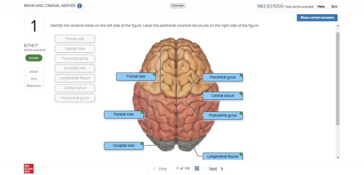
BRAIN AND CRANIAL NERVES BSC 2085 Flashcards | Chegg.com
Solved Exam 2 Homework 55 Correctly label the following - Chegg Biology questions and answers. Exam 2 Homework 55 Correctly label the following meninges of the brain. Meningeal layer Arachnoid mater Dura mater: Pia mater Falx cerebri Arachnoid villus Periosteal layer Subdural space Subarachnoid space 0.89 points. Question: Exam 2 Homework 55 Correctly label the following meninges of the brain.
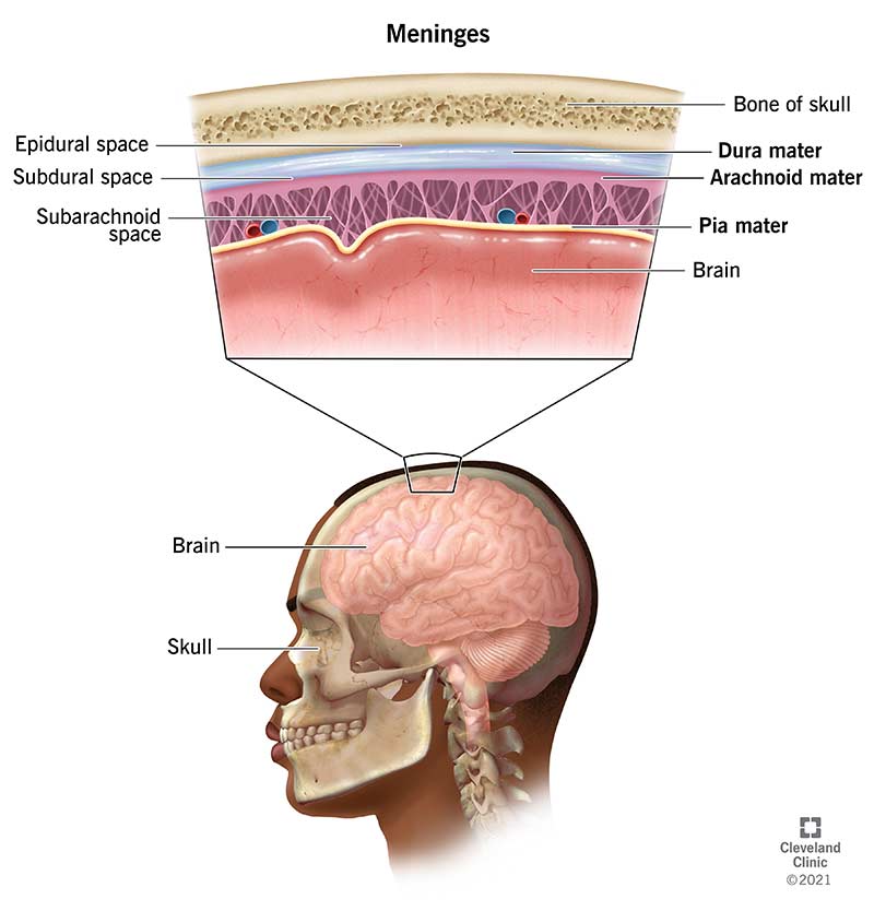
Meninges: What They Are & Function
PDF Brain Review and Wkst Answer - Mayfield City Schools Created Date: 4/30/2013 4:05:46 PM

Chapter 14 Worksheet Flashcards | Quizlet
5 Lobes Of The Brain (A Complete Guide) - NeuroTray 5 lobes of the brain. Each cerebral hemisphere is divided into five lobes: the frontal lobe, the parietal lobe, the occipital lobe and the temporal lobe, four of which have the same name as the bone above them. Deep within the lateral sulcus lies a fifth lobe, the insula or Island of Reil.

17E60840-5AD9-448F-B7D2-E1DB70FD4769.jpeg - Correctly label ...
Positions and Functions of the Four Brain Lobes | MD-Health.com The occipital lobe, the smallest of the four lobes of the brain, is located near the posterior region of the cerebral cortex, near the back of the skull. The occipital lobe is the primary visual processing center of the brain. Here are some other functions of the occipital lobe: Visual-spatial processing. Movement and color recognition.
ANATOMI SISTEM SARAF BIOPSIKOLOGI Unita Werdi Rahajeng - ppt ...
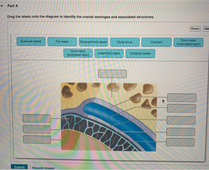
Solved Part A Drag the labels onto the diagram to identify ...
14.3 The Brain and Spinal Cord – Anatomy & Physiology
Trigeminal nerve (CN V): Anatomy, function and branches | Kenhub

Brain - Brain, Spinal Cord, and Nerve Disorders - MSD Manual ...
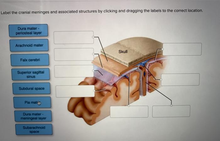
Solved Label the cranial meninges and associated structures ...
Correctly label the following meninges of the brain. | Chegg.com

Solved heducation.com Brain and Spinal Cord Correctly label ...

25B0CC8B-E772-4BFA-B379-ABCDCE963642.jpeg - Correctly label ...

lab 7 (exercise 14) Flashcards | Quizlet

AHCDW10Notes4.pdf - 4. Award: 10.00 points Problems? Adjust ...
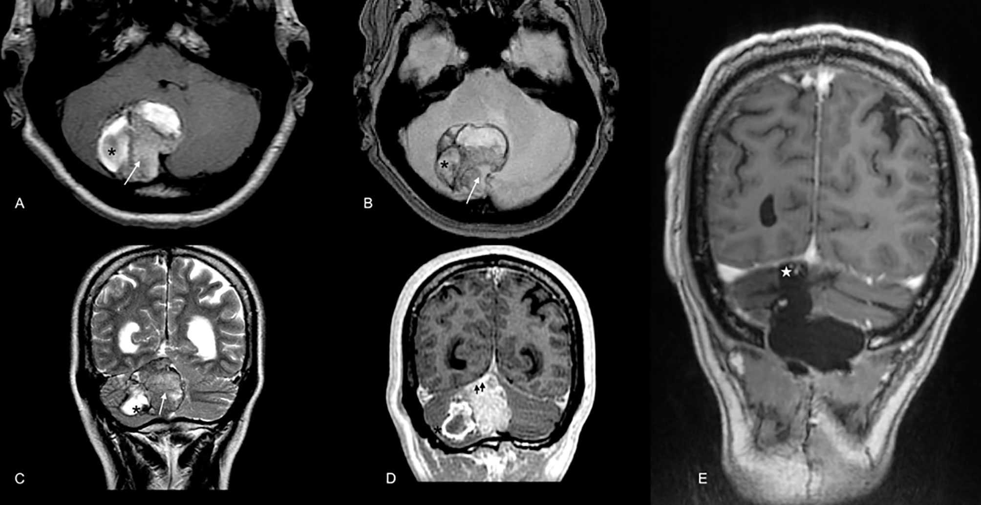
Imaging Modalities: Neuropathology | SpringerLink
Anatomy Exam 2 Flashcards - Easy Notecards

A&P 1 final Flashcards | Quizlet

25B0CC8B-E772-4BFA-B379-ABCDCE963642.jpeg - Correctly label ...

Cerebrum - an overview | ScienceDirect Topics

The correct sequence of meninges from inner to outer side is
Drag the correct label to the appropriate location to label ...
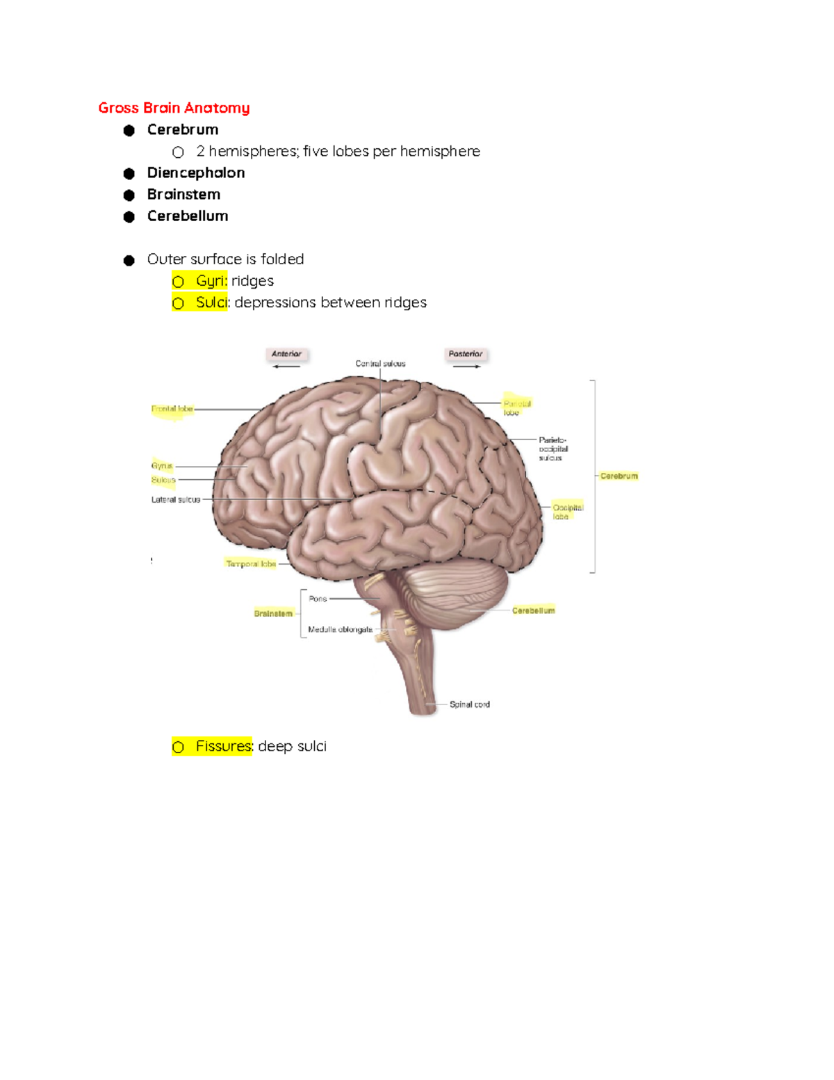
Central Nervous System - Gross Brain Anatomy ○ Cerebrum ○ 2 ...
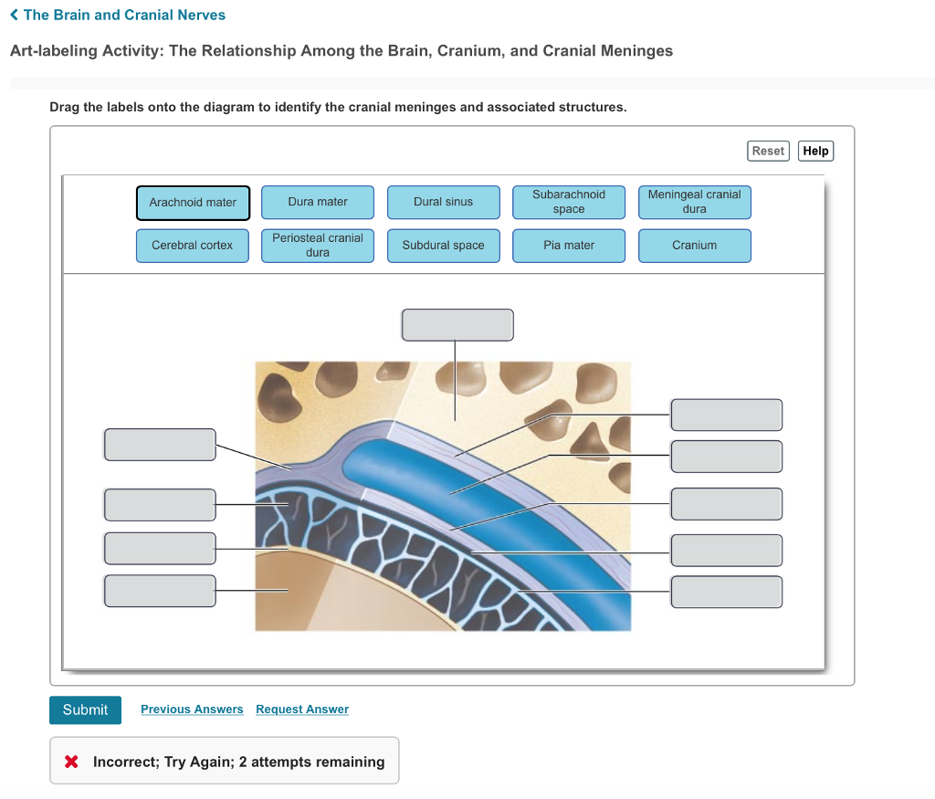
Solved K The Brain and Cranial Nerves Art-labeling Activity ...

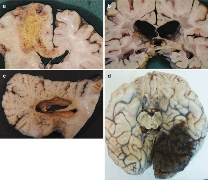
Postmortem examination of patient H.M.'s brain based on ...

QUIZ 7- Meninges of the brain 17.7 part 1 Diagram | Quizlet

Meninges - Wikipedia

The Microbiota-Gut-Brain Axis | Physiological Reviews
Solved Correctly label the following meninges and associated ...

Histology of Central Nervous System Dr. Sama ul Haque. - ppt ...
Meninges, Ventricles, CSF and brain blood supply | Kenhub
Frontiers | Perivascular Spaces, Glymphatic System and MR

CBIO Figures Flashcards | Quizlet
The Human Brain

Meninges of Brain Quiz


Komentar
Posting Komentar