44 diagram of the eye to label
Eye diagram by Firkin - Pinterest Oct 29, 2016 - From a public domain drawing on Wikimedia Commons. Labeled Eye Diagram - Pinterest Labeled Eye Diagram | Best Diagram Collection. Labeled Eye Diagram.
Label Eye Printout - EnchantedLearning.com Label the Eye Diagram. Human Anatomy. Read the definitions, then label the eye anatomy diagram below. Cornea - the clear, dome-shaped tissue covering the front of the eye. Iris - the colored part of the eye - it controls the amount of light that enters the eye by changing the size of the pupil.
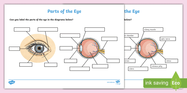
Diagram of the eye to label
Diagram human eye anatomy with label vector image Diagram of human eye anatomy with label illustration. Download a free preview or high-quality Adobe Illustrator (ai), EPS, PDF vectors and high-res JPEG and ... Human Ear Diagram - Bodytomy The Structure of Human Ear. Helix: It is the prominent outer rim of the external ear. Antihelix: It is the cartilage curve that is situated parallel to the helix. Crus of the Helix: It is the landmark of the outer ear, situated right above the pointy protrusion known as the tragus. Auditory Ossicles: The three small bones in the middle ear ... Eye Anatomy: 16 Parts of the Eye & Their Functions - Vision Center The following are parts of the human eyes and their functions: 1. Conjunctiva. The conjunctiva is the membrane covering the sclera (white portion of your eye). The conjunctiva also covers the interior of your eyelids. Conjunctivitis, often known as pink eye, occurs when this thin membrane becomes inflamed or swollen.
Diagram of the eye to label. PDF Eye Anatomy Handout - National Institutes of Health of light entering the eye. Lens: The lens is a clear part of the eye behind the iris that helps to focus light, or an image, on the retina. Macula: The macula is the small, sensitive area of the retina that gives central vision. It is located in the center of the retina. Optic nerve: The optic nerve is the largest sensory nerve of the eye. How to Draw Human Eye Diagram Step by step for beginners How to draw human eye diagram step by step for class 10 student drawing as a beginner by fine arts Guruji with some new and easy technique that helps in boar... Diagram of the Eye Side View No Labels Illustration - Twinkl Sep 18, 2019 - Diagram of the Eye Side View No Labels,Eye,Science,Human Body,Biology,Diagram,Human,Pupil,Sense,Organ,Sclera,Cornea,Iris,Lens,Retina,Optic ... Eye Diagram With Labels and detailed description - BYJUS Well-Labelled Diagram of Eye The anterior chamber of the eye is the space between the cornea and the iris and is filled with a lubricating fluid,... The vascular layer of the eye, known as the choroid contains the connective tissue. The iris and the choroid are connected by the ciliary body. Fovea ...
eye labeling Diagram | Quizlet sclera. Tough white out covering of the eyeball. choroid. Middle layer of the eye (between the retina and the sclera) that contains the blood vessels that nourish the eye and cornea. iris. colored layer that dilates and constricts to allow in more or less light. ciliary body. structure on each side of the lens that connects the choroid and iris. Anatomy of the eye: Quizzes and diagrams | Kenhub Take a look at the diagram of the eyeball above. Here you can see all of the main structures in this area. Spend some time reviewing the name and location of each one, then try to label the eye yourself - without peeking! - using the eye diagram (blank) below. Unlabeled diagram of the eye. Click below to download our free unlabeled diagram of the eye. Structure and Functions of Human Eye with labelled Diagram - BYJUS The External Structure of an Eye. Sclera: It is a white visible portion. It is made up of dense connective tissue and protects the inner parts. Conjunctiva: It lines the sclera and is made up of stratified squamous epithelium. It keeps our eyes moist and clear and provides lubrication by secreting mucus and tears. human eye diagram to label Label the Eye. 9 Pics about Label the Eye : Label the Eye, What Are Eye Dilating Drops? (with pictures) and also Head & Neck Stock Art. Label The Eye . eye diagram blank labeled human anatomy label eyeball drawing labels worksheet quiz coloring purposegames answers parts printable eyes worksheets physiology
Eye labeling Diagram | Quizlet a ring of muscle tissue that forms the colored portion of the eye around the pupil and controls the size of the pupil opening. Cornea. The clear tissue that covers the front of the eye. Posterior Compartment. filled with vitreous humor. Pupil. opening in the center of the iris. Susponsory Ligament. Blank Eye Diagram - Healthiack Best viewed on 1280 x 768 px resolution in any modern browser. Blank eye diagram 1063. Blank eye diagram 1020. Blank eye diagram 1023. Blank eye diagram 1029. Blank eye diagram 1031. Blank eye diagram 1033. Blank eye diagram 1034. Blank eye diagram 1035. labeling the eye worksheet 33 Eye Diagram To Label - Label Design Ideas 2020 dandelionsandthings.blogspot.com. Print Exercise 9: Overview Of The Skeleton: Classification And . structure bones skeleton classification bone overview spongy cartilages exercise which short flat easynotecards. Diagram of eye with labels - simplediagram.netlify.app This is an exercise for students to label a simple blank eye diagram with the following parts. Iris optic nerve pupil cornea lens retina. For us to see there has to be light. When light shines on an object a reflection is sent which passes through the eye lens and later. Labeled Diagram Of An Eye. Oktober 12 2021 Posting Komentar.
FREE! - Label the Eye Worksheet – Teacher-Made Learning Resources
PDF Parts of the Eye - National Institutes of Health Eye Diagram Handout Author: National Eye Health Education Program of the National Eye Institute, National Institutes of Health Subject: Handout illustrating parts of the eye Keywords: parts of the eye, eye diagram, vitreous gel, iris, cornea, pupil, lens, optic nerve, macula, retina Created Date: 12/16/2011 12:39:09 PM
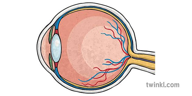
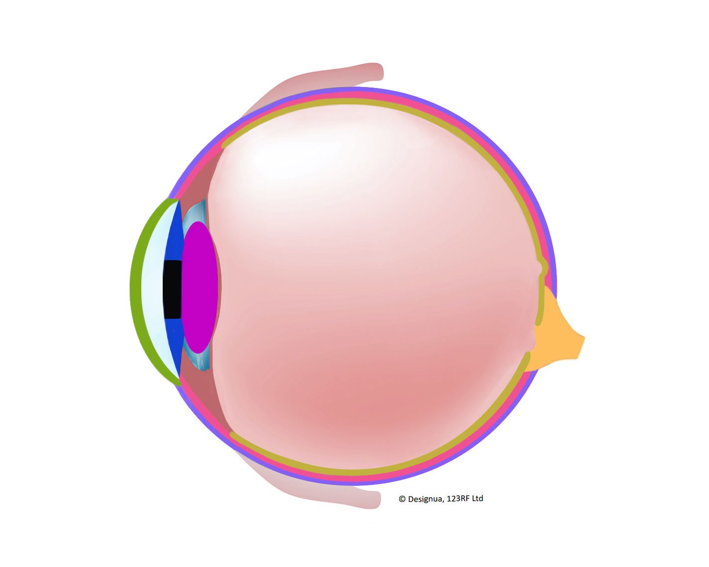
Diagram of the Eye Side View No Labels Illustration - Twinkl
Eye Anatomy Diagram - EnchantedLearning.com Eye Anatomy Diagram Printout. Aqueous humor - the clear, watery fluid inside the eye. It provides nutrients to the eye. Astigmatism - a condition in which the lens is warped, causing images not to focus properly on the retina. Binocular vision - the coordinated use of two eyes which gives the ability to see the world in three dimensions - 3D. Cones - cells the in the retina that sense color.

eye labeling Diagram | Quizlet
What Does the Eye Look Like? - Diagram of the Eye | Harvard Eye Associates It is mostly water and gives the eye its form and shape. Our eyes are vital for seeing the world around us. Keep them healthy by maintaining regular vision exams. Contact Harvard Eye Associates at 949-951-2020 or harvardeye.com to schedule an appointment today.

Diagram of human eye anatomy with label 1945551 Vector Art at ...
Eye Anatomy: Parts of the Eye and How We See Here is a tour of the eye starting from the outside, going in through the front and working to the back. Eye Anatomy: Parts of the Eye Outside the Eyeball. The eye sits in a protective bony socket called the orbit. Six extraocular muscles in the orbit are attached to the eye. These muscles move the eye up and down, side to side, and rotate the eye.

Labeled Eye Diagram | Human eye diagram, Eye anatomy, Diagram ...
Label the Eye Worksheet - Teacher-Made Learning Resources - Twinkl The first page is a labelling exercise with two diagrams of the human eye. One is a view from the outside, and the other is a more detailed cross-section. Challenge learners to label the parts of the eye diagram. On the second page, you'll find a set of answers showing the properly labelled human eyes, designed to help you check the worksheets without having to come up with your own answer key.
Label the Eye
Eye Diagram Quiz - ProProfs Quiz Try this amazing Eye Diagram Quiz quiz which has been attempted 5479 times by avid quiz takers. Also explore over 72 similar quizzes in this category. Take Quizzes. Animal; Nutrition; ... Can you label the parts of the eye in the quiz below? Give it a try and evaluate yourself. The eye has many important parts, each with different functions ...

A schematic diagram of a horizontal section through an eye ...
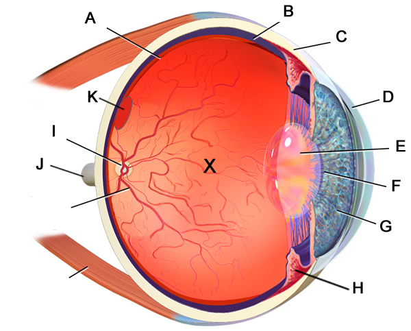
Label Parts of the Human Eye - University of Dayton Parts of the Eye. Select the correct label for each part of the eye. The image is taken from above the left eye. Click on the Score button to see how you did. Incorrect answers will be marked in red. ...

The Eye - Science Quiz
Diagram of the Eye - Lions Eye Institute The eye - one of the most complex organisms in the human body. It is made up of many different parts working in unison together. In order for the eye to work at its best, all parts must work well collectively. To understand the eye and its functions, it's important to understand how the eye works, see below diagrams for both the external ...
/GettyImages-695204442-b9320f82932c49bcac765167b95f4af6.jpg)
Structure and Function of the Human Eye
Module 1: Labeled Diagram of the Eye - Pinterest Module 1: Labeled Diagram of the Eye Itinerary Template, Label Templates, Microsoft Word ... eye anatomy and experiments Science Fair, Science Lessons, ...

Diagram of human eye anatomy with label illustration Stock ...
Label the Eye Quiz - PurposeGames.com This is an online quiz called Label the Eye. There is a printable worksheet available for download here so you can take the quiz with pen and paper. From the quiz author ... Definition And Term Match Up Game For The Eye 12p Image Quiz. Definitions of the Terms of the Skin 12p Multiple-Choice. Playlists by same creator. LegoA1's favorites 2 ...

The diagram above shows the structure of the eye. Choose the ...
Module 1: Labeled Diagram of the Eye - Pinterest Module 1: Labeled Diagram of the Eye Itinerary Template, Label Templates, Microsoft Word ... eye anatomy and experiments Science Fair, Science Lessons, ...

Eye diagram by Firkin | Human eye diagram, Diagram of the eye ...
Labelling the eye - Science Learning Hub In this interactive, you can label parts of the human eye. Use your mouse or finger to hover over a box to highlight the part to be named. Drag and drop the text labels onto the boxes next to the eye diagram. If you want to redo an answer, click on the box and the answer will go back to the top so you can move it to another box.

Pin on Ylli
Labeled Eye Diagram | Eye anatomy diagram, Eye anatomy, Diagram of the eye Pharmacy Images. Medicine Images. This printable contains 13 clear and simple cross sectional diagrams of the human eye. They photocopy well and are great for use as a labeling and coloring exercise for your students. The core eye anatomy diagram, designed as the labeling exercise, has a fully colored and labeled reference ...
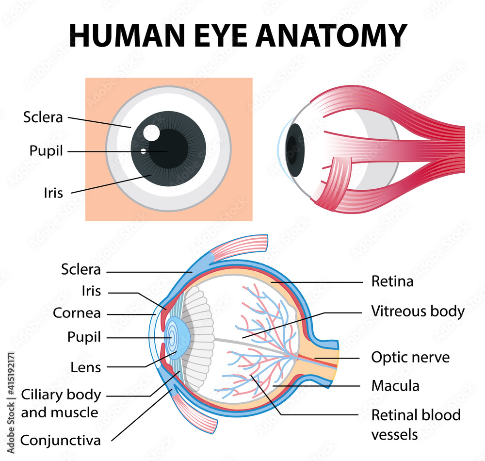
Diagram of human eye anatomy with label Stock Vector | Adobe ...
Easy eye diagram | Labeled eye diagram - Pinterest Dec 7, 2020 - Simple eye diagram | Easy eye diagram | Labeled eye diagram We provide you Simple eye diagram and easy eye diagram from exam point of view.
Diagram Of The Eye For Kids To Label - ClipArt Best
Eye Diagram Printable: Free Worksheet for Kids Completing this worksheet will help your child: • Identify different parts of the eye using an image. • Match the words to the correct part of the eye. • Gain new vocabulary. Help your little learner gain important vocabulary and understand more about their bodies and senses with this quick worksheet about the eye!

Module 1: Labeled Diagram of the Eye | Diagram of the eye ...
Labeled Eye Diagram | Science Trends The iris is a structure found in the eyes of most mammals, and along with the pupil it controls how much light enters the eye. The iris is comprised of two different layers: the stroma, and the pigmented epithelial cells. The upper layer, the stroma, is linked to muscles that contract and dilate the pupil.

Human Eye Diagram | Label Diagram Of Eye | Science Drawing
Labelled Diagram of Human Eye, Explanation and Function - VEDANTU The human eye is a part of the sensory nervous system. Labeled Diagram of Human Eye . The eyes of all mammals consist of a non-image-forming photosensitive ganglion within the retina which receives light, adjusts the dimensions of the pupil, regulates the availability of melatonin hormones, and also entertains the body clock.
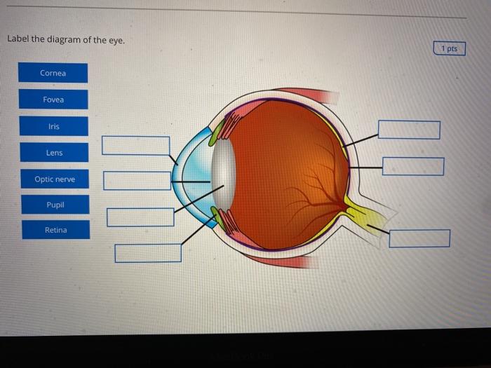
Solved Label the diagram of the eye. 1 pts Cornea Fovea Iris ...
Eye Diagram - Differentiated Worksheets and EASEL ... Jan 29, 2016 - Use these simple eye diagrams to help students learn about the human ... Write the words without a word bank Labels include: eyebrow, eyelid, ...

Label the Eye Worksheet - Homeschool Helper Online | Parts of ...
Labelling the eye — Science Learning Hub The human eye contains structures that allow it to perceive light, movement and colour differences. In this activity, students use online or paper resources to identity and label the main parts of the human eye. Citizen science. Teacher PLD.

Correctly Label the Eye Diagram Quiz
Eye Anatomy: 16 Parts of the Eye & Their Functions - Vision Center The following are parts of the human eyes and their functions: 1. Conjunctiva. The conjunctiva is the membrane covering the sclera (white portion of your eye). The conjunctiva also covers the interior of your eyelids. Conjunctivitis, often known as pink eye, occurs when this thin membrane becomes inflamed or swollen.
:max_bytes(150000):strip_icc()/GettyImages-695204442-b9320f82932c49bcac765167b95f4af6.jpg)
Structure and Function of the Human Eye
Human Ear Diagram - Bodytomy The Structure of Human Ear. Helix: It is the prominent outer rim of the external ear. Antihelix: It is the cartilage curve that is situated parallel to the helix. Crus of the Helix: It is the landmark of the outer ear, situated right above the pointy protrusion known as the tragus. Auditory Ossicles: The three small bones in the middle ear ...
Eye With Labels Clip Art at Clker.com - vector clip art ...
Diagram human eye anatomy with label vector image Diagram of human eye anatomy with label illustration. Download a free preview or high-quality Adobe Illustrator (ai), EPS, PDF vectors and high-res JPEG and ...
Eye Lesson

Solved: Label the diagram. Refer to Figure 43-18 to check ...
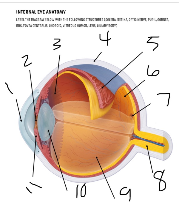
Solved INTERNAL EYE ANATOMY LABEL THE DIAGRAM BELOW WITH THE ...

Sensory Structures | BioNinja

Diagram and label the internal structures of the eye, and gi ...

Diagram of Human Eye Anatomy with Label Stock Vector ...

Label the Eye Quiz

Diagram human eye anatomy with label Royalty Free Vector

Cross Section of a Human Eye Diagram Black and White ...
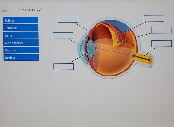
Solved Label the parts of the eye. Sclera Choroid Lens Optic ...

human eye label diagram | How to draw eye diagram easily | Eyeball diagram drawing | eye anatomy
Labelling the eye - Teaching resources
Changing the way you learn | Quiz

Quick Quiz
Diagram of the Eye - Lions Eye Institute

a Draw a labelled diagram of the human eye. Label the ...
Structures of the Eye
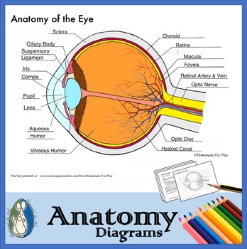
Anatomy of the Eye Diagrams for Coloring/Labeling, with Reference and Summary

KLB Science Interactivities - The Human Eye
Label Eye Printout - EnchantedLearning.com
Eye Anatomy Diagram - EnchantedLearning.com
Labelling the eye — Science Learning Hub


Komentar
Posting Komentar