39 light microscope diagram with labels
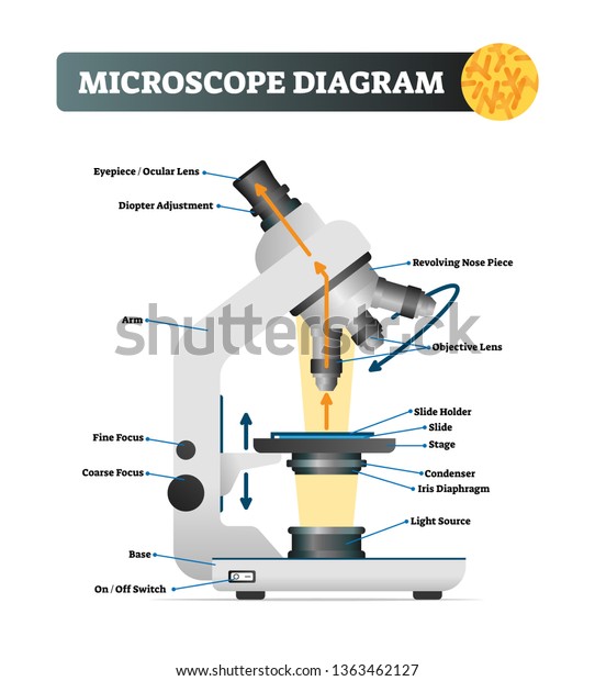
Label the microscope Diagram | Quizlet Diaphragm. Regulates the amount of light on the specimen. Light Source. Projects light upwards through the diaphragm, the specimen, and the lenses. Arm. supports the body tube. Stage. Supports the slide being viewed. Coarse Adjustment. Label Microscope Diagram - EnchantedLearning.com low-power objective - a small lens with low magnifying power. mirror (or light source) - this directs light upwards onto the slide. revolving nosepiece - the rotating device that holds the objectives (lenses). stage - the platform on which a slide is placed. stage clips - metal clips that hold a slide securely onto the stage.
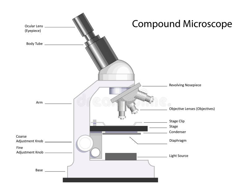
Compound Microscope Parts, Functions, and Labeled Diagram Nov 18, 2020 · Parts of a Compound Microscope Each part of the compound microscope serves its own unique function, with each being important to the function of the scope as a whole. The individual parts of a compound microscope can vary heavily depending on the configuration & applications that the scope is being used for. Common compound microscope parts include: Compound Microscope Definitions for ...

Light microscope diagram with labels
Microscope, Microscope Parts, Labeled Diagram, and Functions Revolving Nosepiece or Turret: Turret is the part of the microscope that holds two or multiple objective lenses and helps to rotate objective lenses and also helps to easily change power. Objective Lenses: Three are 3 or 4 objective lenses on a microscope. The objective lenses almost always consist of 4x, 10x, 40x and 100x powers. The most common eyepiece lens is 10x and when it coupled with ... Compound Microscope Parts - Labeled Diagram and their Functions The eyepiece (or ocular lens) is the lens part at the top of a microscope that the viewer looks through. The standard eyepiece has a magnification of 10x. You may exchange with an optional eyepiece ranging from 5x - 30x. [In this figure] The structure inside an eyepiece. The current design of the eyepiece is no longer a single convex lens. Simple Microscope - Diagram (Parts labelled), Principle, Formula and Uses Parts of a Simple Microscope A simple microscope consists of Optical parts Mechanical parts Labeled Diagram of simple microscope parts Optical parts The optical parts of a simple microscope include Lens Mirror Eyepiece Lens A simple microscope uses biconvex lens to magnify the image of a specimen under focus.
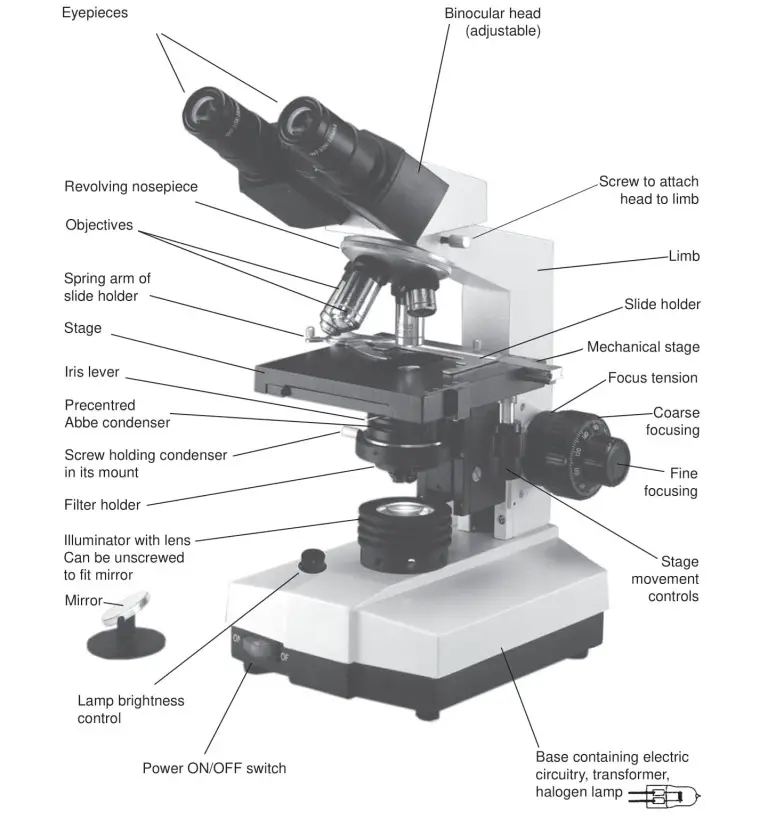
Light microscope diagram with labels. Label Microscope Diagram - EnchantedLearning.com low-power objective - a small lens with low magnifying power. mirror (or light source) - this directs light upwards onto the slide. revolving nosepiece - the rotating device that holds the objectives (lenses). stage - the platform on which a slide is placed. stage clips - metal clips that hold a slide securely onto the stage. Binocular Microscope Anatomy - Parts and Functions with a Labeled Diagram Now, I will discuss the details anatomy of the light compound microscope with the labeled diagram. Why it is called binocular: because it has two ocular lenses or an eyepiece on the head that attaches to the objective lens, this ocular lens magnifies the image produced by the objective lens. Binocular microscope parts and functions Find Jobs in Germany: Job Search - Expat Guide to Germany ... Browse our listings to find jobs in Germany for expats, including jobs for English speakers or those in your native language. Parts of a microscope with functions and labeled diagram - Microbe Notes Figure: Diagram of parts of a microscope There are three structural parts of the microscope i.e. head, base, and arm. Head - This is also known as the body. It carries the optical parts in the upper part of the microscope. Base - It acts as microscopes support. It also carries microscopic illuminators.
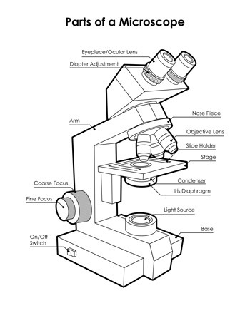
Compound Microscope - Diagram (Parts labelled), Principle and Uses See: Labeled Diagram showing differences between compound and simple microscope parts Structural Components The three structural components include 1. Head This is the upper part of the microscope that houses the optical parts 2. Arm This part connects the head with the base and provides stability to the microscope. Label the Light Microscope - Labelled diagram Eyepiece, Light Source, Base, Stage, Stage Clips, Fine Focus, Coarse Focus, Arm, Objective Lens, Diaphragm. Label the microscope — Science Learning Hub Jun 08, 2018 · All microscopes share features in common. In this interactive, you can label the different parts of a microscope. Use this with the Microscope parts activity to help students identify and label the main parts of a microscope and then describe their functions. Drag and drop the text labels onto the microscope diagram. If you want to redo an ... Microscope Parts, Function, & Labeled Diagram - slidingmotion Microscope parts labeled diagram gives us all the information about its parts and their position in the microscope. Microscope Parts Labeled Diagram The principle of the Microscope gives you an exact reason to use it. It works on the 3 principles. Magnification Resolving Power Numerical Aperture. Parts of Microscope Head Base Arm Eyepiece Lens
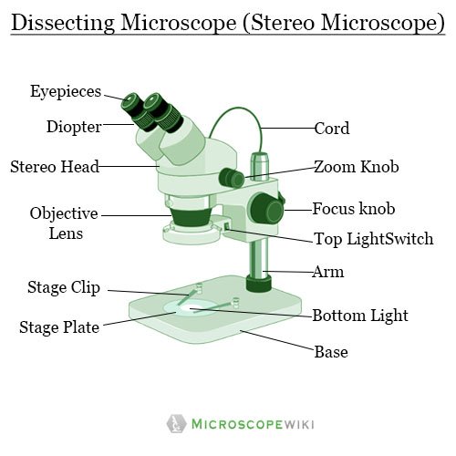
Parts of Stereo Microscope (Dissecting microscope) – labeled ... Labeled part diagram of a stereo microscope Major structural parts of a stereo microscope. There are three major structural parts of a stereo microscope. The viewing Head includes the upper part of the microscope, which houses the most critical optical components, including the eyepiece, objective lens, and light source of the microscope. label microscope worksheet Microscope parts worksheet label diagram light answers labeling middle worksheeto blank compound labeled via label microscope worksheet. 31 Label A Microscope Worksheet - Labels Design Ideas 2020. 15 Pictures about 31 Label A Microscope Worksheet - Labels Design Ideas 2020 : 13 Best Images of Definition Matching Worksheet - Huntington's Disease ... PDF Parts of the Light Microscope - Science Spot Supports the MICROSCOPE D. STAGE CLIPS HOLD the slide in place C. OBJECTIVE LENSES Magnification ranges from 10 X to 40 X F. LIGHT SOURCE Projects light UPWARDS through the diaphragm, the SPECIMEN, and the LENSES H. DIAPHRAGM Regulates the amount of LIGHT on the specimen E. STAGE Supports the SLIDE being viewed K. ARM Used to SUPPORT the Microscope Labeling - The Biology Corner 1) Start with scanning (the shortest objective) and only use the COARSE knob . Once it is focused… 2) Switch to low power (medium) and only use the COARSE knob . You may need to recenter your slide. Once it is focused.. 3) Switch to high power (long objective).
Simple Microscope - Parts, Functions, Diagram and Labelling ...
Fluorescence Resonance Energy Transfer (FRET) Microscopy Presented in Figure 3 is a Jablonski diagram illustrating the coupled transitions involved between the donor emission and acceptor absorbance in fluorescence resonance energy transfer. Absorption and emission transitions are represented by straight vertical arrows (green and red, respectively), while vibrational relaxation is indicated by wavy ...
Vektor Stok Microscope Diagram Vector Illustration Labeled ...
Parts of a Microscope Labeling Activity - Storyboard That Knowing the names of the different parts of the microscope is essential to be able to use one properly. Create a poster that labels the parts of a microscope and includes descriptions of what each part does. Click "Start Assignment". Use a landscape poster layout (large or small). Search for a diagram of a microscope.

Dissecting Stereo Microscope Parts and Functions
(PDF) Introduction to Microscopy - ResearchGate Nov 08, 2017 · 1. Microscopy with light and electrons 2. Electron/specimen interactions: processes and detectors 3. The electron microscope family 4. Specimen preparation for electron microscopy 5.

Microscope labeled diagram
Microscope Diagram - Label Diagram | Quizlet The bottom of the microscope, used for support. ocular lens. Eyepiece of a microscope. Diaphragm. Regulates the amount of light on the specimen. nosepiece of microscope. holds the objective lenses. objective lens. The lens on a light microscope that is closest to the stage.

Parts of a microscope with functions and labeled diagram
Join LiveJournal Password requirements: 6 to 30 characters long; ASCII characters only (characters found on a standard US keyboard); must contain at least 4 different symbols;

Microscope - diagram Tom Butler | Microscope parts, Science ...
Labelled Diagram Of A Light Microscope - GlobalSpec A schematic diagram for the microscope -based label -free microfluidic light scattering cytometer. IB Biology/Option H - Further Human Physiology - Wikibooks, open books for an open world Draw and label a diagram showing a transverse section of the ileum as seen under a light microscope .

The Science Break - Labels for the light microscope for GCSE ...
Bright-field microscope (Compound light microscope) - Diagram (Parts ... Bright-field Microscope. A bright-field microscope, also known as a compound light microscope is among the simplest of optical microscopes. Optical microscopes employ visible light and a series of lenses to magnify the specimen and view it in detail. A bright-field microscope uses light rays to create a dark image against a bright background ...
Dissecting microscope (Stereoscopic or stereo microscope ...
Labeling the Parts of the Microscope | Microscope World Resources Labeling the Parts of the Microscope This activity has been designed for use in homes and schools. Each microscope layout (both blank and the version with answers) are available as PDF downloads. You can view a more in-depth review of each part of the microscope here. Download the Label the Parts of the Microscope PDF printable version here.
Compound Microscope stock vector. Illustration of research ...
Fluorescence - Wikipedia Fluorescence is the emission of light by a substance that has absorbed light or other electromagnetic radiation.It is a form of luminescence.In most cases, the emitted light has a longer wavelength, and therefore a lower photon energy, than the absorbed radiation.A perceptible example of fluorescence occurs when the absorbed radiation is in the ultraviolet …
Free Microscope Drawing, Download Free Microscope Drawing png ...
Light Microscope- Definition, Principle, Types, Parts, Labeled Diagram ... A light microscope is a biology laboratory instrument or tool, that uses visible light to detect and magnify very small objects and enlarge them. They use lenses to focus light on the specimen, magnifying it thus producing an image. The specimen is normally placed close to the microscopic lens.

Free Microscope Drawing, Download Free Microscope Drawing png ...
Microscope Types (with labeled diagrams) and Functions Simple microscope labeled diagram Simple microscope functions It is used in industrial applications like: Watchmakers to assemble watches Cloth industry to count the number of threads or fibers in a cloth Jewelers to examine the finer parts of jewelry Miniature artists to examine and build their work Also used to inspect finer details on products

Microscope - Label - Part 2 Diagram | Quizlet
Light microscopes - Cell structure - Edexcel - BBC Bitesize The components of a light microscope and their functions Calculating the magnification of light microscopes. The compound microscope uses two lenses to magnify the specimen: the eyepiece and an ...

Microscope Parts and Functions
16 Parts of a Compound Microscope: Diagrams and Video Once you have an understanding of the parts of the microscope it will be much easier to navigate around and begin observing your specimen, which is the fun part! The 16 core parts of a compound microscope are: Head (Body) Arm. Base. Eyepiece. Eyepiece tube.

Microscope Labeling
A Study of the Microscope and its Functions With a Labeled Diagram ... These labeled microscope diagrams and the functions of its various parts, attempt to simplify the microscope for you. However, as the saying goes, 'practice makes perfect', here is a blank compound microscope diagram and blank electron microscope diagram to label. Download the diagrams and practice labeling the different parts of these ...

Microscope diagram labeled | Clipart Panda - Free Clipart Images
Microscope Parts and Functions With Labeled Diagram and Functions How does a Compound Microscope Work? Before exploring microscope parts and functions, you should probably understand that the compound light microscope is more complicated than just a microscope with more than one lens. Parts of a Chainsaw - Anatomy Explained [2022 Update]

Label Microscope Diagram - EnchantedLearning.com
PDF Label the Light Microscope - New Paltz Middle School Label the Light Microscope: Ocular lens Objective ourse Adjustment Fine Adjustment Light ase Diaphragm rightness control Nosepiece ondenser Mechanical Stage Head with prism Ocular tube Arm. Head Nosepiece Objective turret Stage Iris Diaphragm Illumination System Base Eyepiece ...

MICROSCOPE Labeling - Part - 3
Simple Microscope - Diagram (Parts labelled), Principle, Formula and Uses Parts of a Simple Microscope A simple microscope consists of Optical parts Mechanical parts Labeled Diagram of simple microscope parts Optical parts The optical parts of a simple microscope include Lens Mirror Eyepiece Lens A simple microscope uses biconvex lens to magnify the image of a specimen under focus.

A Study of the Microscope and its Functions With a Labeled ...
Compound Microscope Parts - Labeled Diagram and their Functions The eyepiece (or ocular lens) is the lens part at the top of a microscope that the viewer looks through. The standard eyepiece has a magnification of 10x. You may exchange with an optional eyepiece ranging from 5x - 30x. [In this figure] The structure inside an eyepiece. The current design of the eyepiece is no longer a single convex lens.

The Compound Light Microscope Diagram | Quizlet
Microscope, Microscope Parts, Labeled Diagram, and Functions Revolving Nosepiece or Turret: Turret is the part of the microscope that holds two or multiple objective lenses and helps to rotate objective lenses and also helps to easily change power. Objective Lenses: Three are 3 or 4 objective lenses on a microscope. The objective lenses almost always consist of 4x, 10x, 40x and 100x powers. The most common eyepiece lens is 10x and when it coupled with ...

Parts of a Microscope - SmartSchool Systems

Types of Microscopes: Definition, Working Principle, Diagram ...

Compound Microscope- Definition, Labeled Diagram, Principle ...

Parts Of A Microscope Labeling Teaching Resources | TpT

Label the microscope — Science Learning Hub
Parts of a Microscope with Their Functions – Microbe Online
Labeled Microscope Diagram – Tim's Printables

easy compound microscope diagram - Clip Art Library
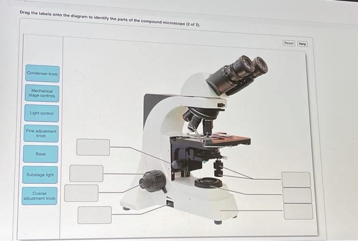
Solved Drag the labels onto the diagram to identify the ...

Microscope Diagram Labeled, Unlabeled and Blank | Parts of a ...

This is a common compound microscope. Label its parts from A ...

Microscope, Microscope Parts, Labeled Diagram, and Functions
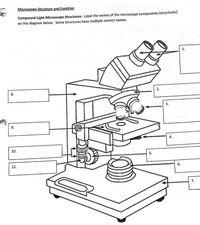
Answered: Microscope Structure and Function… | bartleby

Compound Microscope – Diagram (Parts labelled), Principle and ...

Microscope With Labels clip art | Microscope parts ...

Microscope Maintenance Tips | Science supplies, Multi step ...

Microscope With Labels Clip Art at Clker.com - vector clip ...

Compound Microscope Parts, Diagram Definition, Application ...

Labelled Microscope with Functions | Microscope parts ...

Parts of Stereo Microscope (Dissecting microscope) – labeled ...


Komentar
Posting Komentar